


₹ 5,799 ₹ 15,157

₹ 17,499 ₹ 32,975

₹ 8,699 ₹ 22,379

₹ 1,599 ₹ 4,955

₹ 6,399 ₹ 15,039

₹ 1,499 ₹ 4,955

₹ 1,699 ₹ 4,955

₹ 5,999 ₹ 7,645

₹ 5,299 ₹ 8,011
 - Myaskro")
₹ 8,999 ₹ 22,379

₹ 1,499 ₹ 4,955

₹ 1,699 ₹ 4,955

₹ 7,999 ₹ 22,379
 With Ligaments")
₹ 1,699 ₹ 4,955

₹ 5,499 ₹ 10,201
")
₹ 1,699 ₹ 4,561
 - MYASKRO")
₹ 25,999 ₹ 40,119

₹ 8,199 ₹ 22,379
 With Special Lamination")
₹ 1,599 ₹ 3,865
 | Clinical-Grade Wall Charts - Myaskro")
₹ 6,399 ₹ 8,006
 With Special Lamination")
₹ 1,599 ₹ 3,865

₹ 11,799 ₹ 22,379

₹ 2,199 ₹ 6,957

₹ 1,599 ₹ 4,537
 With Special Lamination")
₹ 1,599 ₹ 3,865

₹ 9,599 ₹ 17,305

₹ 4,199 ₹ 9,316

₹ 5,999 ₹ 8,945

₹ 20,999 ₹ 27,655
")
₹ 6,599 ₹ 8,945

₹ 5,999 ₹ 8,965
")
₹ 6,399 ₹ 8,675

₹ 5,199 ₹ 10,579
")
₹ 7,799 ₹ 12,845
%20educational%20bundle%20-10x10.jpg "Osteoarthritis (OA) Educational Bundle | Knee & Spine 4 stage Degeneration Models + Spine Disorders & Knee Injuries Charts - Myaskro")
₹ 11,599 ₹ 22,379

₹ 9,599 ₹ 16,765

₹ 12,199 ₹ 22,379
 | 52cm x 70cm with Special Lamination")
₹ 6,399 ₹ 18,965

₹ 33,899 ₹ 69,579

₹ 6,799 ₹ 17,659

₹ 7,999 ₹ 14,750

₹ 33,599 ₹ 57,779

₹ 3,999 ₹ 7,315

₹ 12,599 ₹ 25,836
 | Myaskro")
₹ 3,199 ₹ 4,865
")
₹ 9,499 ₹ 17,845

₹ 5,999 ₹ 8,945

₹ 11,799 ₹ 18,945
")
₹ 169,299 ₹ 184,965
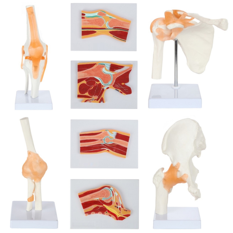
MYASKRO Anatomical Joints & Cross-Sections Model Bundle (8 Models)
A complete ortho-teaching kit pairing four life-size joints with four matching cross-section boards. Perfect for MBBS/BPT/BOT programs, orthopedic labs, and OSCE/OSPE stations.
Key Benefits
Two views per region: 3D joint + sectional slab for shoulder, elbow, knee and hip/pelvis—instant correlation of osteology to soft-tissue planes.
Clinically precise detail: Capsules, key ligaments, articular cartilage, labrum/menisci, bursae, and surrounding myofascial layers.
Biomechanics made visual: Compare hinge vs ball-and-socket motion, end-feel, and stability constraints; map common injury mechanisms.
Exam & viva ready: Ideal for demonstrating special tests (Lachman, valgus/varus, apprehension/relocation, impingement concepts, FABER).
Built for classrooms: Stable bases, wipe-clean surfaces, high-contrast colour coding for repeated handling.
Eight-model bundle: shoulder, elbow, knee and hip joints with matching cross-sections to teach ligaments, capsules, labrum/menisci and biomechanics—made for colleges and skill labs.
This MYASKRO bundle delivers comprehensive limb anatomy by combining macro 3D joints with sectional context.
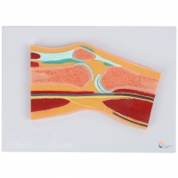
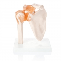
Shoulder (glenohumeral): Glenoid labrum, capsule, coracoacromial/coracoclavicular and glenohumeral ligaments; section shows subacromial space, cuff and neurovascular corridors for impingement/instability discussions.
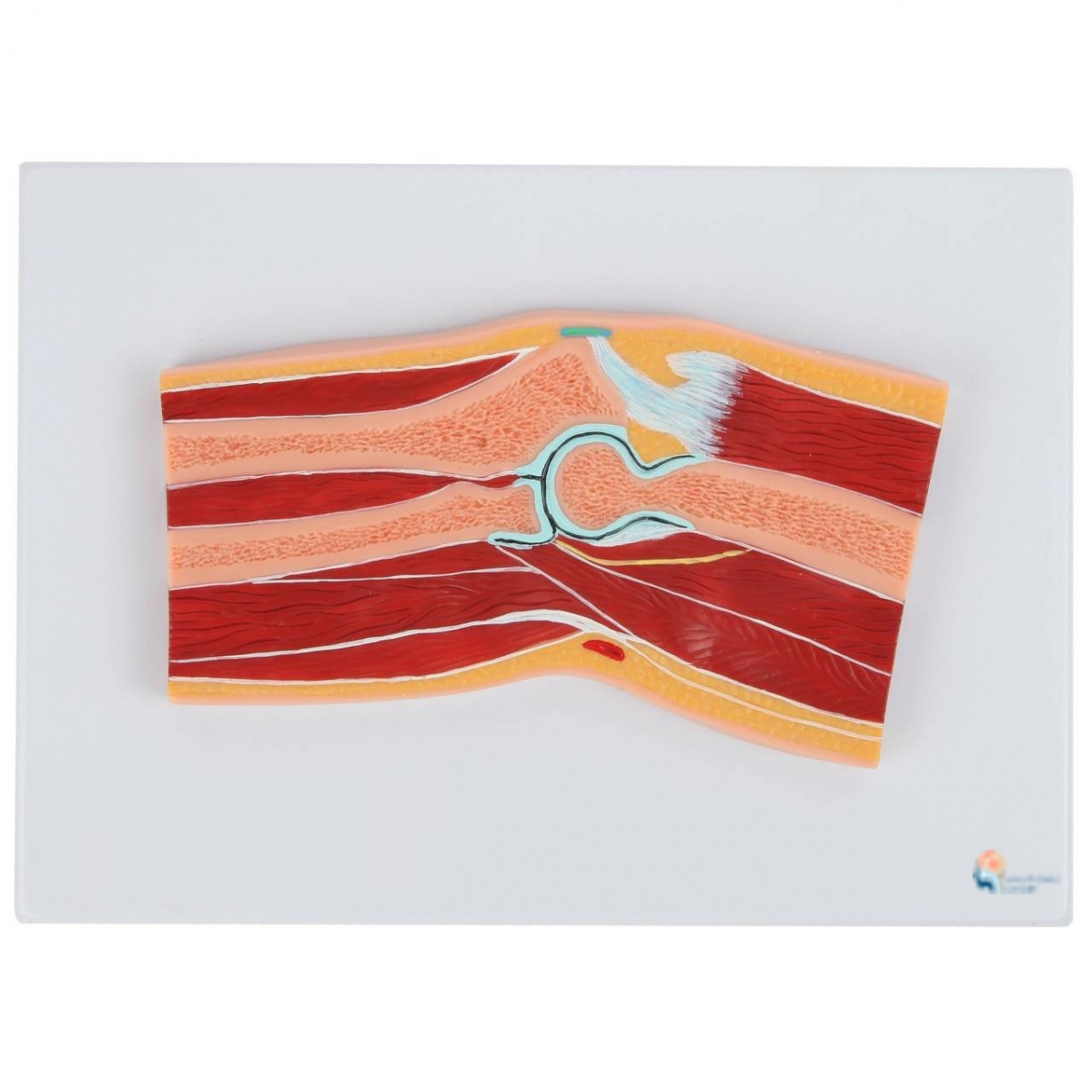
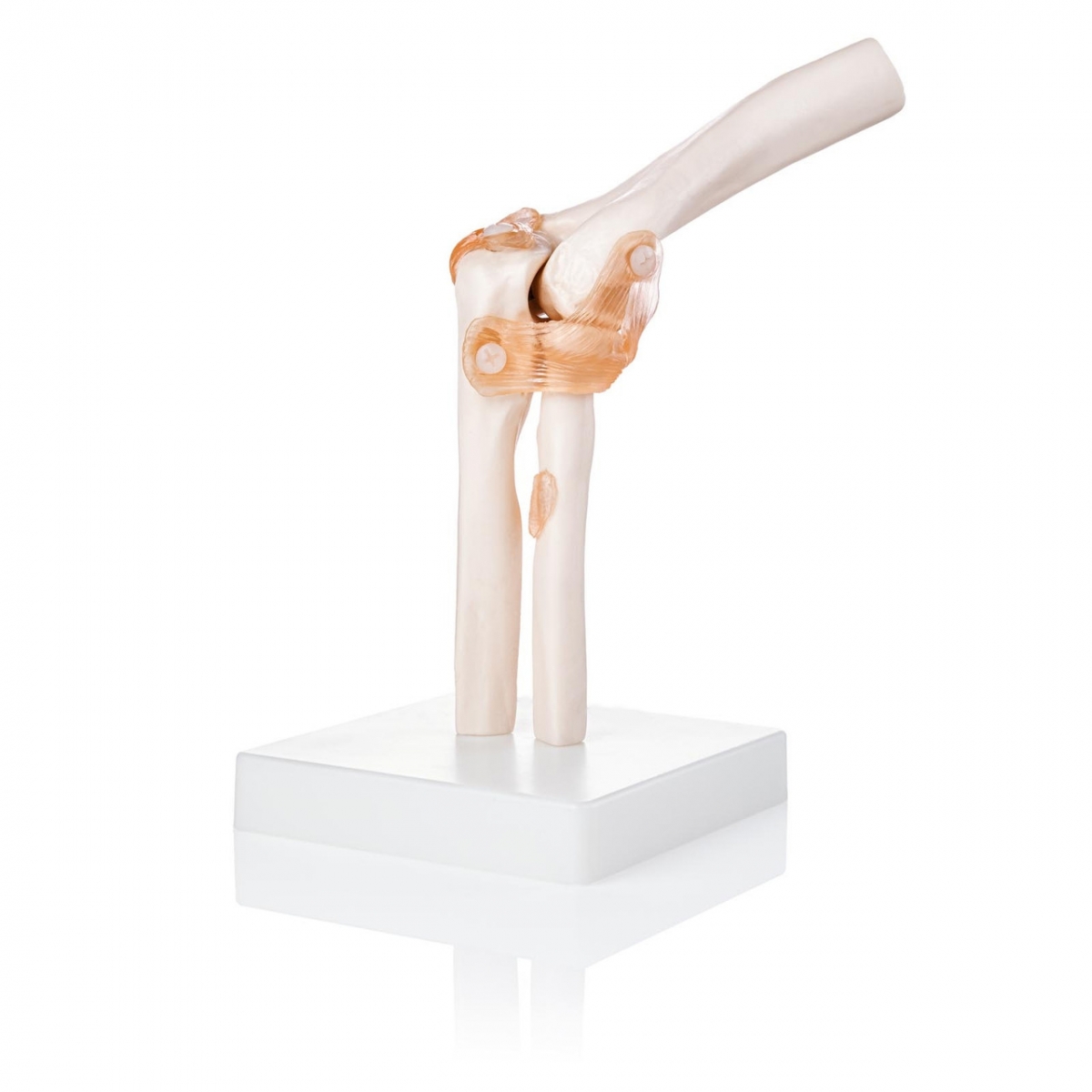
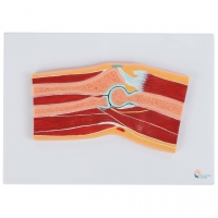
Elbow: Ulnar/radial collateral and annular ligaments with humero-ulnar/humero-radial alignment; section highlights flexor–extensor compartments and cubital tunnel relations.
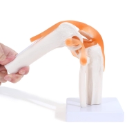
Knee: ACL, PCL, MCL, LCL, patellar ligament and meniscal contours; section demonstrates tibiofemoral/ patellofemoral relations for OA/meniscal and instability concepts.
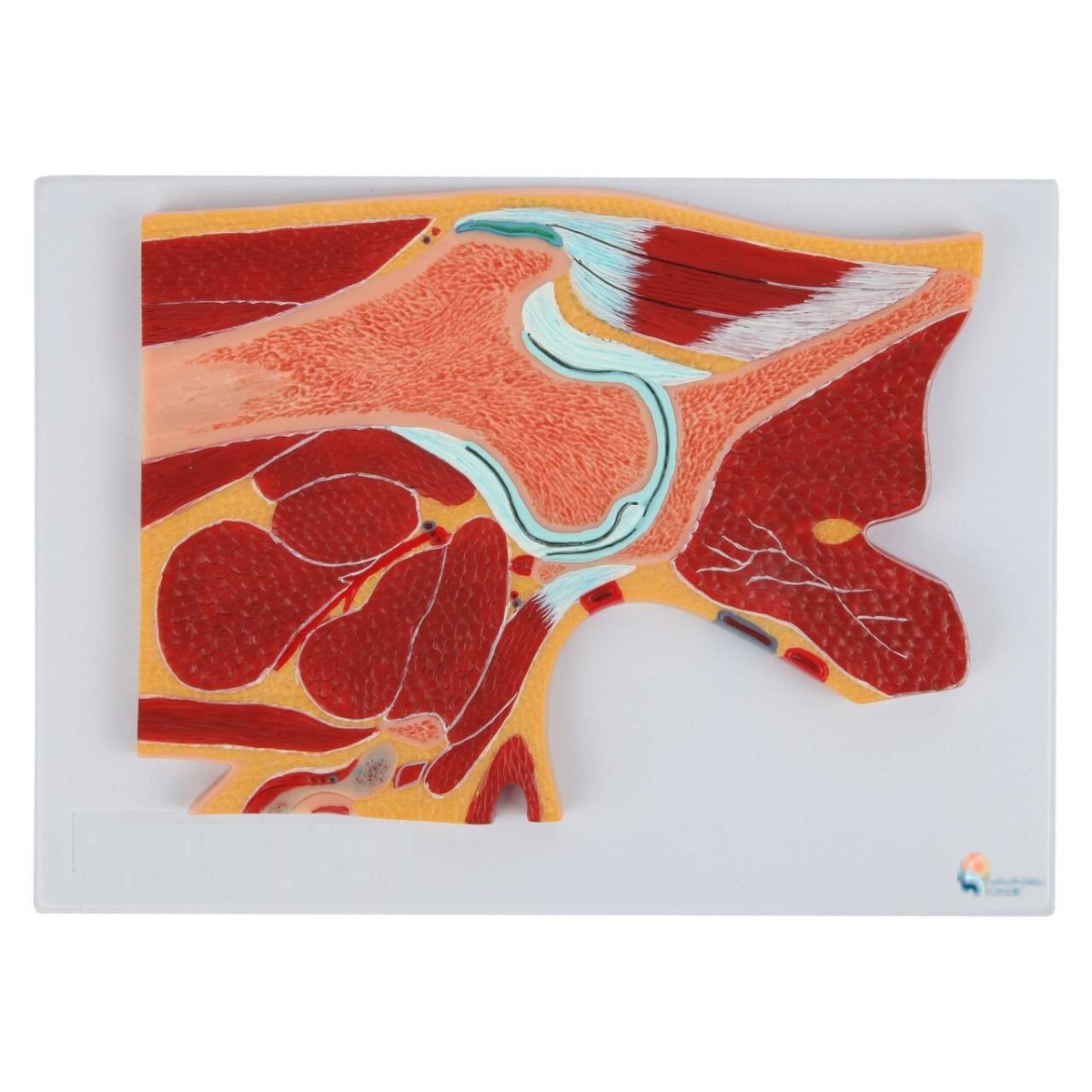
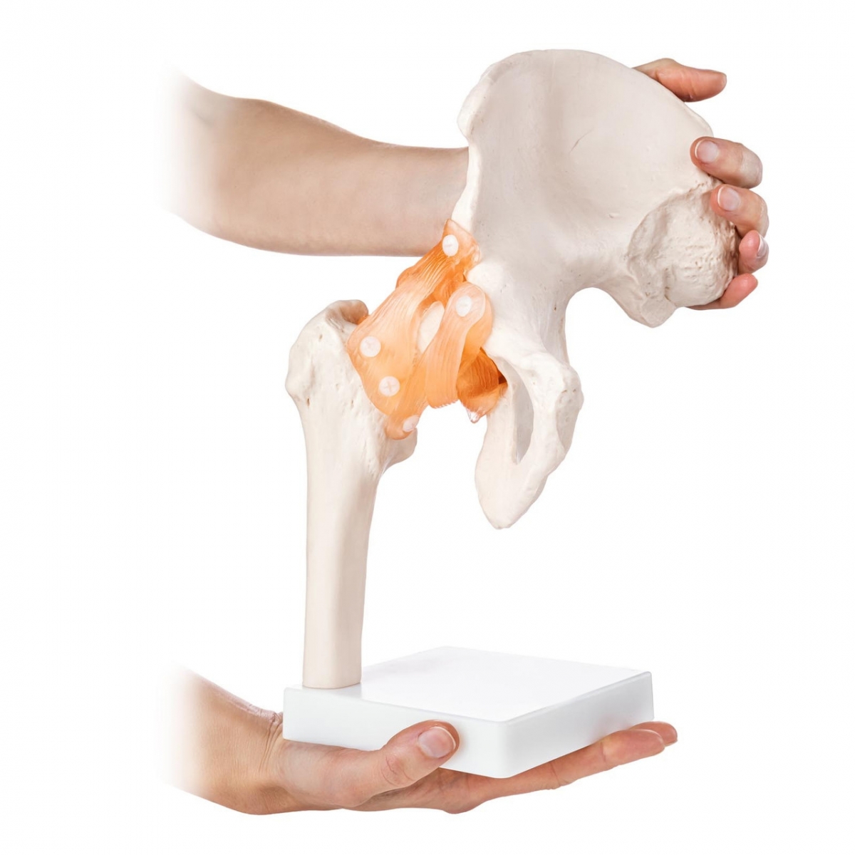
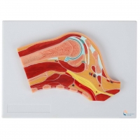
Hip/Pelvis: Iliofemoral, pubofemoral, ischiofemoral ligaments, acetabular labrum and cartilage; section maps capsule, gluteal/iliopsoas planes and neurovascular paths, supporting FAI and OA teaching.
Durable materials withstand high-throughput lab use and routine disinfection.
Teaching Objectives & Skills Practised
Identify bones, capsules and named ligaments for shoulder, elbow, knee and hip.
Explain joint classification, axes of motion and stability mechanisms.
Correlate sectional planes with X-ray/MRI and surface anatomy.
Relate injury mechanisms to clinical tests (education only).
Structure OSCE/OSPE stations using accurate landmarks and movement demos.
What’s Included
1 × Shoulder Joint Section Anatomy Model
1 × Knee Joint Section Anatomy Model
1 × Elbow Joint Section Anatomy Model
1 × Pelvic (Hip) Joint Section Anatomy Model
1 × Shoulder Joint Model (life size)
1 × Knee Joint Model (life size)
1 × Elbow Joint Model (life size)
1 × Hip Joint Model (life size)