

₹ 3,999 ₹ 7,315

₹ 33,899 ₹ 69,579
%20educational%20bundle%20-10x10.jpg "Osteoarthritis (OA) Educational Bundle | Knee & Spine 4 stage Degeneration Models + Spine Disorders & Knee Injuries Charts - Myaskro")
₹ 11,599 ₹ 22,379

₹ 4,199 ₹ 10,555

₹ 11,799 ₹ 22,379

₹ 7,999 ₹ 14,750

₹ 8,199 ₹ 22,379

₹ 20,599 ₹ 28,965

₹ 5,999 ₹ 8,945

₹ 11,799 ₹ 18,945
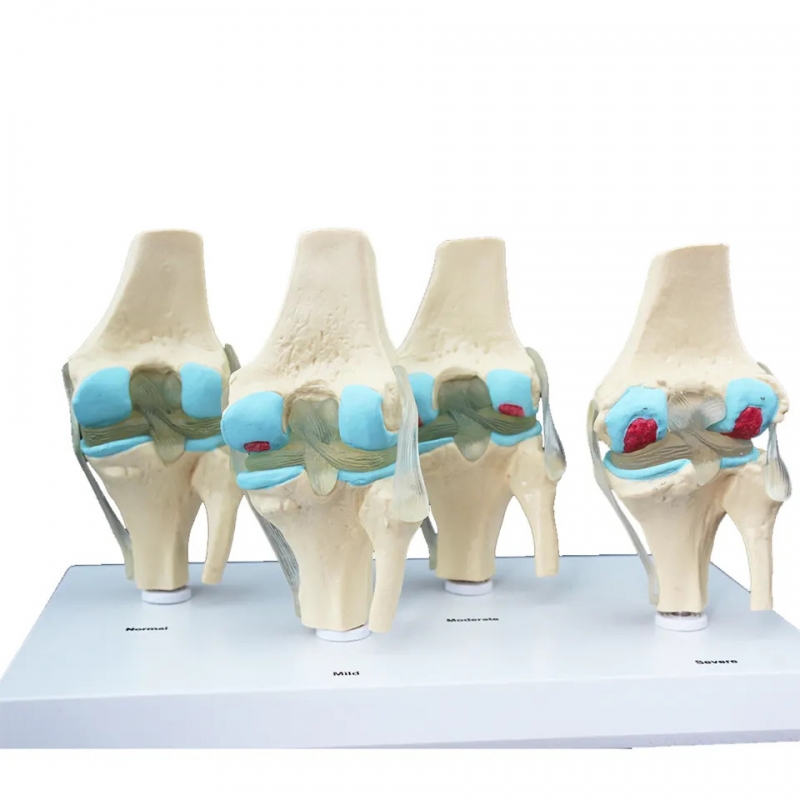
Understanding arthritis isn't just about diagnosis—it's about visualising the progressive damage that occurs within the knee joint. The MYASKRO Knee 4-Stage Arthritis Model gives you the perfect educational platform to show the difference between a healthy knee and degenerative osteoarthritis across four clearly defined stages: Normal, Mild, Moderate, and Severe.
Whether you are an orthopedic specialist, physiotherapist, rheumatologist, or anatomy educator, this model allows you to demonstrate the exact anatomical changes that accompany knee arthritis—including cartilage erosion, synovial thickening, osteophyte formation, joint space narrowing, and inflammatory swelling.
The model consists of four life-size knee joint replicas mounted on a shared base. Each model is color-coded and labelled to indicate:
Crafted from durable, medical-grade PVC, the model is ideal for frequent use in clinics, classrooms, and consultation rooms. It helps simplify complex diagnoses for patients and enhance understanding for medical trainees.
This is especially useful in India’s orthopedic and physiotherapy ecosystem where patient education and conservative joint care are priorities. Whether you’re showing an elderly patient why their pain is worsening or training a batch of BPT interns on degenerative joint conditions—this tool gives you the visual backup you need.
“Using this model at Sanjay Gandhi Institute, Lucknow has made our joint clinics smoother. Patients immediately understand where they fall on the arthritis scale,” shares Dr. Aditi Rawat, Orthopedic Faculty.
For educators and clinicians who deal with knee pain daily, this 4-stage arthritis model is the missing link between scans, symptoms, and understanding.
The MYASKRO 4-Stage Knee Arthritis Model isn’t just informative—it’s transformative in how clearly it communicates the reality of knee degeneration. Designed for hands-on use in orthopedic, physiotherapy, and academic environments, it illustrates the step-by-step breakdown of joint health—from full function to advanced deterioration.
Each of the four knee replicas shows progressive pathological changes that occur with osteoarthritis:
This model supports clinical explanations, classroom instruction, and patient decision-making. Use it to:
Made from non-toxic, premium PVC plastic, the knees are textured and color-coded to highlight cartilage, ligaments, bone, and inflammatory areas. Each model is permanently affixed to a clean base with printed labels—ideal for professional tabletop use in hospitals, colleges, and diagnostic centres.
“I use this daily at my ortho OPD in CMC Vellore. Explaining treatment timelines based on this model is far more effective than just showing MRI reports,” says Dr. Karan Shetty, Senior Consultant Orthopedic Surgeon.
Designed for repetitive teaching and consultation use, this model does what no chart or scan can—it shows patients and learners the journey of joint damage in clear, tactile form.
When it comes to osteoarthritis, visuals speak louder than reports. The MYASKRO Knee 4-Stage Arthritis Model gives professionals and educators the ability to show—rather than just describe—what’s happening inside the joint. And that makes all the difference when it comes to patient trust, student engagement, and clinical communication.
This model doesn't rely on generic diagrams or vague descriptions. It presents the knee in its true form, stage by stage, showing the gradual breakdown of cartilage, narrowing of joint space, and emergence of structural deformities. Each knee is meticulously designed to depict real-world pathology in a way that both novices and professionals can understand instantly.
For practitioners, it enhances your ability to explain why a treatment plan varies for early-stage arthritis versus advanced degeneration. For teachers, it becomes a hands-on asset for practical sessions, viva prep, or orthopedic module lectures. For patients, it builds clarity—and with that comes better decision-making.
The compact footprint, durable build, and clean base make it easy to set up anywhere—from OPD desks and seminar tables to rehabilitation stations and anatomy labs. Whether you're conducting a CME session, doing rounds, or holding a joint camp, this model performs with quiet precision.
It’s already used across top-tier institutions and clinics in India, helping bridge the gap between symptoms and solutions. This isn’t just a model—it’s a daily tool in the hands of those who diagnose, explain, and heal.
“We now use it in every pre-surgery counselling session. Patients feel more informed and confident after seeing the exact condition replicated in the model,” says Dr. Rajat Verma, Ortho Dept., Narayana Hospital, Jaipur.
Some teaching aids show structure. Others show progression. This one shows both—and does it with the kind of detail that turns confusion into understanding. When explaining joint degeneration, there’s no better ally at your side than this model. It helps you bring the invisible into view—and that’s where real impact begins.
Total Reviews (0)