


₹ 12,599 ₹ 25,836

₹ 7,999 ₹ 22,379

₹ 20,999 ₹ 27,655

₹ 5,999 ₹ 8,965
 With Special Lamination")
₹ 1,599 ₹ 3,865
 With Special Lamination")
₹ 1,599 ₹ 3,865

₹ 3,999 ₹ 7,315

₹ 1,599 ₹ 4,955

₹ 17,499 ₹ 32,975

₹ 5,799 ₹ 15,157

₹ 6,399 ₹ 15,039

₹ 1,699 ₹ 4,955

₹ 1,499 ₹ 4,955

₹ 5,299 ₹ 8,011
 - Myaskro")
₹ 8,999 ₹ 22,379

₹ 5,499 ₹ 10,201
")
₹ 1,699 ₹ 4,561
 - MYASKRO")
₹ 25,999 ₹ 40,119

₹ 1,599 ₹ 4,537

₹ 11,799 ₹ 22,379

₹ 4,199 ₹ 9,316

₹ 9,599 ₹ 17,305
%20educational%20bundle%20-10x10.jpg "Osteoarthritis (OA) Educational Bundle | Knee & Spine 4 stage Degeneration Models + Spine Disorders & Knee Injuries Charts - Myaskro")
₹ 11,599 ₹ 22,379

₹ 5,199 ₹ 10,579

₹ 12,199 ₹ 22,379

₹ 33,899 ₹ 69,579
")
₹ 6,599 ₹ 8,945
")
₹ 43,899 ₹ 58,965

₹ 20,599 ₹ 28,965
 | Myaskro")
₹ 3,199 ₹ 4,865
")
₹ 6,399 ₹ 8,945

₹ 11,799 ₹ 18,945
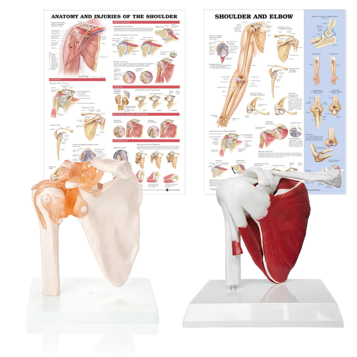
Components
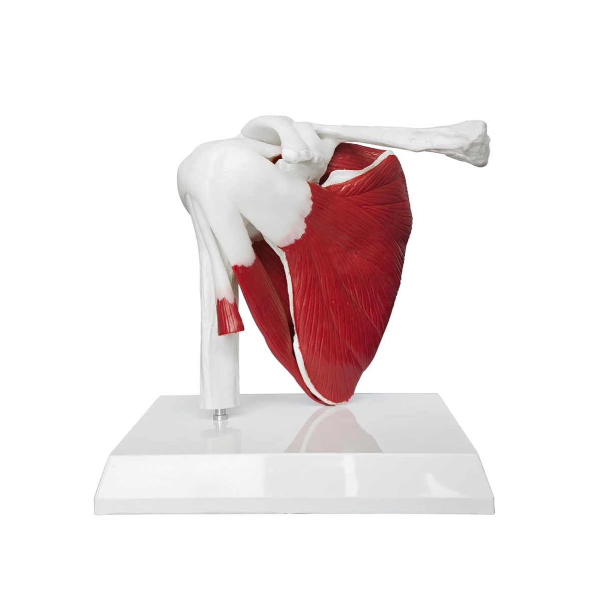
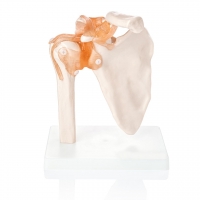
Muscular Shoulder Joint Model: Humerus–scapula–clavicle with superficial myology of the rotator cuff (supraspinatus, infraspinatus, teres minor, subscapularis) and deltoid regions; visible long head biceps tendon and glenoid labrum.
Shoulder Joint Model with Ligaments: Capsuloligamentous complex including superior/middle/inferior glenohumeral ligaments, coracoacromial, acromioclavicular, coracoclavicular (conoid, trapezoid), and transverse humeral ligaments; articulated for demonstration of physiologic ranges.
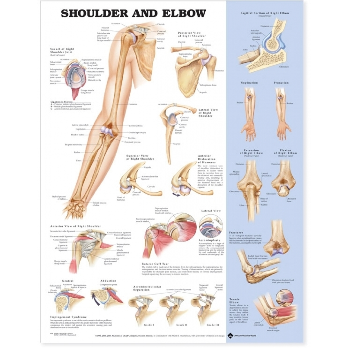
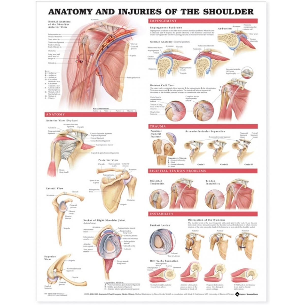
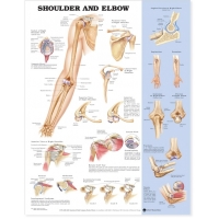
Charts (52 × 70 cm, special lamination with rollers):
Anatomy of the Shoulder (osseous, capsular, myologic maps)
Injuries of the Shoulder (impingement, rotator-cuff tear, SLAP/Bankart lesions, dislocation, AC sprain, adhesive capsulitis)
Learning Objectives
Identify osseous landmarks: acromion, coracoid, glenoid, greater/lesser tubercle, bicipital groove.
Correlate rotator-cuff function with dynamic stability and humeral head centering.
Explain capsuloligamentous restraint across abduction/external rotation; relate to anterior instability.
Map common pathologies to anatomic structures: impingement (subacromial), cuff tears, SLAP/Bankart, AC injuries, frozen shoulder.
Demonstrate scapulohumeral rhythm and planes of motion for bedside/OSCE teaching.
Specifications
Scale: life-size; material: rigid PVC with color-coded structures.
Mounting: individual base stands for each model; 360° viewing.
Charts: heavy-gauge lamination with top–bottom rollers; marker-wipeable.
Use
UG/PG anatomy, orthopaedics, sports medicine, physiotherapy skill labs; patient education for surgical consent and rehabilitation counseling.
Total Reviews (0)