


₹ 20,999 ₹ 27,655

₹ 5,199 ₹ 10,579

₹ 1,599 ₹ 4,537

₹ 33,899 ₹ 69,579

₹ 9,599 ₹ 17,305

₹ 6,399 ₹ 15,039

₹ 7,950 ₹ 15,299
")
₹ 1,699 ₹ 4,561
 With Ligaments")
₹ 1,699 ₹ 4,955

₹ 1,599 ₹ 4,955

₹ 1,699 ₹ 4,955

₹ 8,499 ₹ 22,445

₹ 5,799 ₹ 15,157

₹ 17,499 ₹ 32,975
 With Special Rigid Lamination and Plastic Rollers - Myaskro")
₹ 1,599 ₹ 3,265

₹ 7,999 ₹ 14,750

₹ 11,499 ₹ 12,865

₹ 20,599 ₹ 28,965

₹ 11,799 ₹ 18,945
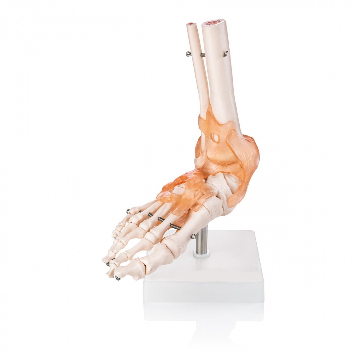
Understanding the human foot means more than just knowing the bones—it means visualizing how ligaments stabilize, how muscles contract, and how every structure interacts during motion. The Foot Joint Model with Muscles and Ligaments brings this understanding to life, offering a comprehensive, three-dimensional view of one of the most complex weight-bearing structures in the body.
This life-size model integrates both the skeletal framework and soft tissue anatomy of the foot and ankle. You’ll see clearly defined tendons, ligaments, joint capsules, and muscle attachments—all in vivid, color-coded detail. It's an essential asset for medical educators, orthopedic professionals, physiotherapists, and sports rehabilitation trainers.
The model includes articulated representations of the tibia and fibula, connected realistically to the talus, calcaneus, navicular, cuboid, metatarsals, and phalanges. But what sets this apart is the presence of key anatomical structures like the Achilles tendon, plantar fascia, and lateral/medial ligament complexes. You can show not just where pain is—but why it’s there.
Whether you’re demonstrating foot biomechanics in a classroom, explaining surgical procedures in a clinic, or guiding patients through post-injury recovery, this model offers a complete narrative of the foot’s structure and function in motion.
Healthcare providers often ask: “Can it show real mechanical relationships?” Yes—because this model isn’t just a static skeleton. It reveals how muscles stabilize joints, how ligaments restrict motion, and how imbalance leads to dysfunction. It’s an educational tool and a clinical explainer all in one.
Semantic queries like “foot model with ligaments,” “muscle and tendon foot anatomy model,” “3D foot biomechanics tool,” and “anatomical teaching model with soft tissue” all align perfectly with what this model delivers.
“This is the only model I’ve used that shows soft tissue so clearly. It’s a game-changer for both teaching and patient education,” says Prof. Ravi Menon, Orthopedic Trainer, Bangalore.
From sports injuries to chronic foot pain, this model helps visualize causes, demonstrate corrections, and build better understanding from the ground up—literally.
The Foot Joint Model with Muscles and Ligaments is more than a skeletal reference—it’s a multi-layered, functional anatomy model that gives you the full picture of how the foot performs under pressure. This model merges form and function by showing not only the bones of the foot but also key connective tissue structures responsible for stability and movement.
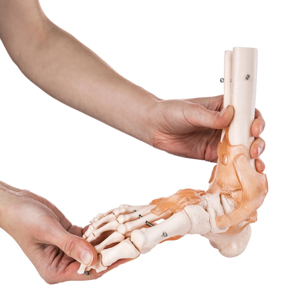
You’ll find clearly detailed Achilles tendon insertion, deltoid ligament, lateral collateral ligaments, plantar fascia, and extensor tendons that traverse from the ankle to the toes. The tendons are shown with natural curvature and anatomical origin-insertion points, allowing for an accurate depiction of mechanical relationships within the joint.
This model is ideal for use in sports medicine, physiotherapy training, orthopedic surgery explanation, podiatry, and academic institutions. It’s particularly effective in demonstrating how overuse, trauma, or misalignment can impact ligaments and muscle-tendon junctions—leading to injuries like plantar fasciitis, sprains, or tendonitis.
Practitioners often ask: “Can I use this to explain injury mechanisms to patients?” Yes. The transparent layout of ligaments and soft tissue makes it easy to identify points of strain or injury. Use it to highlight improper foot biomechanics or demonstrate corrective strategies in post-op care, rehab, or athletic training.
Durability is built into every detail. The soft-tissue components are molded in high-resilience, medical-grade polymer that mimics the look and feel of actual tissue. The skeletal base is cast from high-density, wear-resistant resin, securely mounted on a metal rod and white display base for all-angle visibility.
This model also supports popular search terms such as “foot ligaments model,” “muscle anatomy of foot,” “sports injury teaching aid,” and “anatomical foot model for podiatry.” Because whether online or in a consultation room, it delivers clarity, credibility, and context.
“We use it to demonstrate tendon function and injury risk in our biomechanics lab. Students find it invaluable for understanding real-world motion,” says Laura S., Sports Science Faculty, Dublin.
This model doesn’t just show where structures are—it demonstrates how they work together. And that makes it an essential tool in any anatomy-focused environment.
The foot is a marvel of biological engineering—33 joints, over 100 muscles, tendons, and ligaments, all working in synchrony. To truly understand or explain this complexity, you need more than just a bone model. The Foot Joint Model with Muscles and Ligaments delivers that missing layer of functional insight, offering a holistic view of foot structure and motion.
For students, it transforms dry textbook diagrams into a tactile experience. For clinicians, it becomes a reliable point of reference for explaining pain, instability, and injury mechanisms. And for patients, it answers the most important question: “What’s going on inside my foot?”
It’s perfectly suited for multi-disciplinary use. Whether in a sports rehab facility, university lab, orthopedic clinic, or podiatry practice, the combination of skeletal framework and soft tissue anatomy makes it one of the most versatile tools in anatomical education.
Its structural clarity supports a wide range of learning objectives—from identifying ligament strain patterns to discussing tendon overuse injuries or demonstrating how insoles and braces support proper alignment. Its realistic texture and color scheme make the model intuitive and easy to understand, even for non-medical audiences.
Professionals often wonder, “Does this help patients visualize their condition better?” Absolutely. Seeing the ligaments and tendons in context makes explanations tangible, trustworthy, and easier to accept. Whether you're discussing a rupture, sprain, or inflammation, it reinforces the diagnosis with visual authority.
This model ranks exceptionally well for queries like “3D foot muscle model,” “anatomical foot with tendons and ligaments,” “foot joint soft tissue trainer,” and “clinical foot model for teaching.” It's precisely the kind of tool that both search engines and professionals recognize as valuable.
“It’s a staple in our clinical demonstrations. You can teach mechanics, identify risk factors, and walk patients through recovery—all using one model,” says Dr. Eliot R., Orthopedic Specialist, Chicago.
In the world of foot education, this model stands tall. Quietly accurate. Uncompromisingly detailed. And built to support every step of your teaching, treatment, or training journey—down to the last ligament.
Total Reviews (0)