


₹ 12,599 ₹ 25,836

₹ 33,899 ₹ 69,579
 - Myaskro")
₹ 8,999 ₹ 22,379

₹ 6,399 ₹ 15,326

₹ 5,199 ₹ 10,579

₹ 1,599 ₹ 4,537
")
₹ 1,699 ₹ 4,561
 With Ligaments")
₹ 1,699 ₹ 4,955

₹ 1,599 ₹ 4,955

₹ 1,699 ₹ 4,955

₹ 8,499 ₹ 22,445

₹ 5,799 ₹ 15,157

₹ 17,499 ₹ 32,975

₹ 11,499 ₹ 12,865

₹ 20,599 ₹ 28,965

₹ 11,799 ₹ 18,945
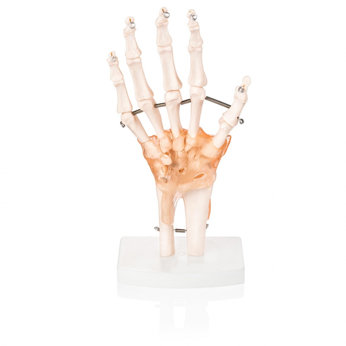
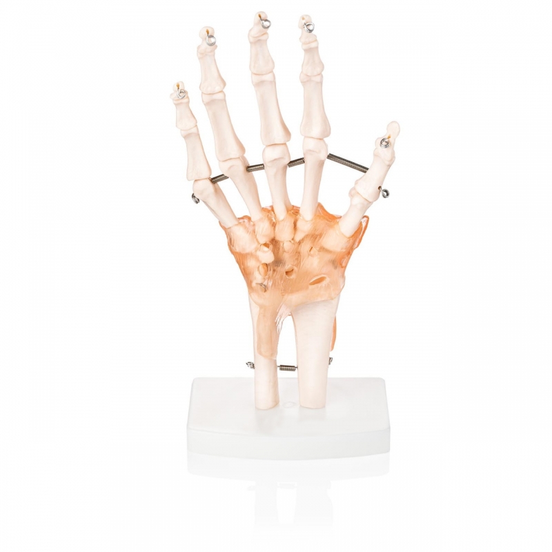
The hand is a masterpiece of anatomical engineering—29 bones, 34 muscles, and dozens of tendons and ligaments working in perfect harmony. The MYASKRO Premium Hand Joint Model with Ligaments captures this complexity in stunning, life-size detail, making it the ideal choice for orthopedics, physiotherapy, anatomy education, and surgical demonstration.
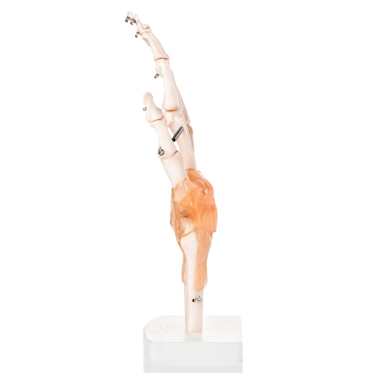
This model features all major bones of the human hand—carpals, metacarpals, phalanges, and distal radius/ulna—held together by semi-flexible ligament structures that mirror real-life anatomical alignment and movement. Mounted on a sturdy white base, it stands upright and is ready for both classroom and clinical display.
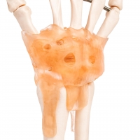
Crafted from non-toxic, medical-grade PVC, the model offers excellent durability and anatomical realism. The ligaments—colored and textured for clarity—allow you to demonstrate joint articulation, ligamentous support, and biomechanical constraints in a highly visual way.
It's widely used by medical professors, hand surgeons, physiotherapists, chiropractic professionals, and rehabilitation educators. Whether you're teaching joint kinematics or explaining a post-injury recovery plan to a patient, this hand model transforms passive listeners into active learners.
Curious about its practical range? This model is perfect for demonstrating:
It ranks strongly for search terms like “hand joint anatomical model,” “ligament hand skeleton model,” “PVC hand with ligaments,” and “orthopedic hand teaching tool.” Those who search for realism and reliability end up right here.
“During our upper limb dissection module at SMS Medical College, Jaipur, this model helped me understand ligament pathways better than any diagram ever could,” says Ritika Verma, MBBS Student.
Compact, detailed, and built to last, this model isn't just a reference—it's a revelation for anyone serious about understanding hand anatomy.
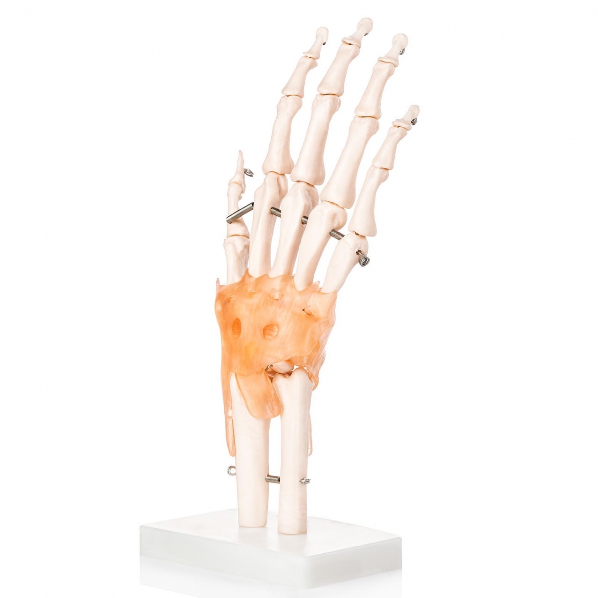
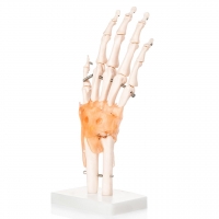
The MYASKRO Hand Joint Model delivers a level of anatomical detail that goes far beyond typical skeletal displays. This isn’t just a static representation—it’s a dynamic educational tool that captures the true mechanical relationships between bones, ligaments, and joints.
Each component is expertly rendered:
The ligaments provide limited flexibility, enabling instructors and clinicians to demonstrate joint constraints, finger extension/flexion paths, and overall range of motion limitations. It's ideal for showing both normal mechanics and pathological conditions like trigger finger, rheumatoid deformity, or ligament laxity.
This model shines in:
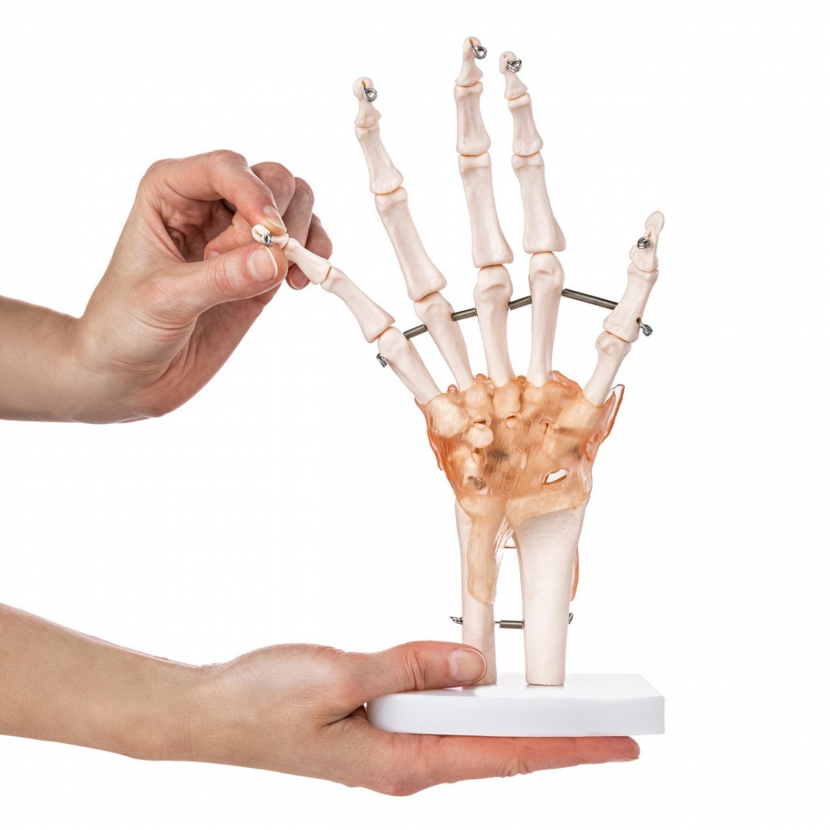
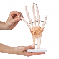
Medical professionals often ask: “Does it include joint movement?” While the bones are held in place for accuracy, the ligament design allows slight mobility and stretch, making it possible to mimic finger movements, joint deviations, and alignment issues in a controlled, instructive way.
Constructed from medical-grade PVC and durable metal fittings, the model is built for years of hands-on use. The mounted base offers stability for demonstrations while keeping the structure upright and visible from multiple angles.
Highly relevant for search terms such as “hand skeleton with ligaments,” “flexible anatomical hand model,” “orthopedic hand demo,” and “hand joint structure teaching tool.”
“In our physio OPD at AIIMS Bhopal, this model helps us explain wrist mobility limitations to patients recovering from fractures and tendon injuries,” shares Dr. Nitin Rao, MPT.
Designed to replicate. Built to educate. Trusted in every setting where precision meets performance.
The human hand is a marvel of biomechanics—capable of fine motor control, strength, and flexibility. But to truly teach or explain its complexity, you need more than illustrations. You need a model that communicates structure, movement, and function with total clarity. The MYASKRO Hand Joint Model with Ligaments does exactly that.
This model isn’t just a reference piece—it’s a functional anatomical guide for those who work with hands every day. Whether you’re a student navigating the intricacies of the carpal tunnel, a physiotherapist guiding a post-op patient, or a surgeon discussing ligament reconstruction, this tool supports your explanations with visual authority.
It allows you to demonstrate conditions such as ligament sprains, osteoarthritis, nerve entrapment syndromes, and range-of-motion deficits. It adds precision to your diagnosis, confidence to your consultation, and effectiveness to your teaching.
Still wondering if it holds up under frequent use? Absolutely. The reinforced joint connections and synthetic ligaments are built for repeated interaction. Use it in rounds, classroom discussions, rehab planning, or public health demos—it’s stable, portable, and made to last.
Search terms like “hand anatomy model with ligaments,” “PVC hand joint for ortho,” “clinical hand model India,” and “functional wrist and finger model” all land right here. And they should. Because this model delivers on every expectation—quality, accuracy, utility, and presentation.
“We recently introduced this in our Occupational Therapy Lab at PGIMER Chandigarh. It’s made our practical training sessions so much more interactive and clear,” says Ms. Aparna Sinha, OT Educator.
The best tools don’t just show what’s there—they explain how it works. This hand joint model offers that unique blend of visual realism and educational depth that professionals rely on daily. A must-have for any setting where anatomy isn’t just studied—it’s applied.
Total Reviews (0)