


")
₹ 1,699 ₹ 4,561

₹ 12,599 ₹ 25,836

₹ 7,999 ₹ 14,750

₹ 33,599 ₹ 57,779

₹ 5,199 ₹ 10,579

₹ 12,199 ₹ 22,379

₹ 9,599 ₹ 16,765
 | Clinical-Grade Wall Charts - Myaskro")
₹ 6,399 ₹ 8,006

₹ 33,899 ₹ 69,579
 With Special Lamination")
₹ 1,599 ₹ 3,865
 With Special Lamination")
₹ 1,599 ₹ 3,865

₹ 5,799 ₹ 15,157

₹ 5,499 ₹ 10,201

₹ 3,999 ₹ 7,315

₹ 20,999 ₹ 27,655

₹ 20,599 ₹ 28,965

₹ 11,799 ₹ 18,945
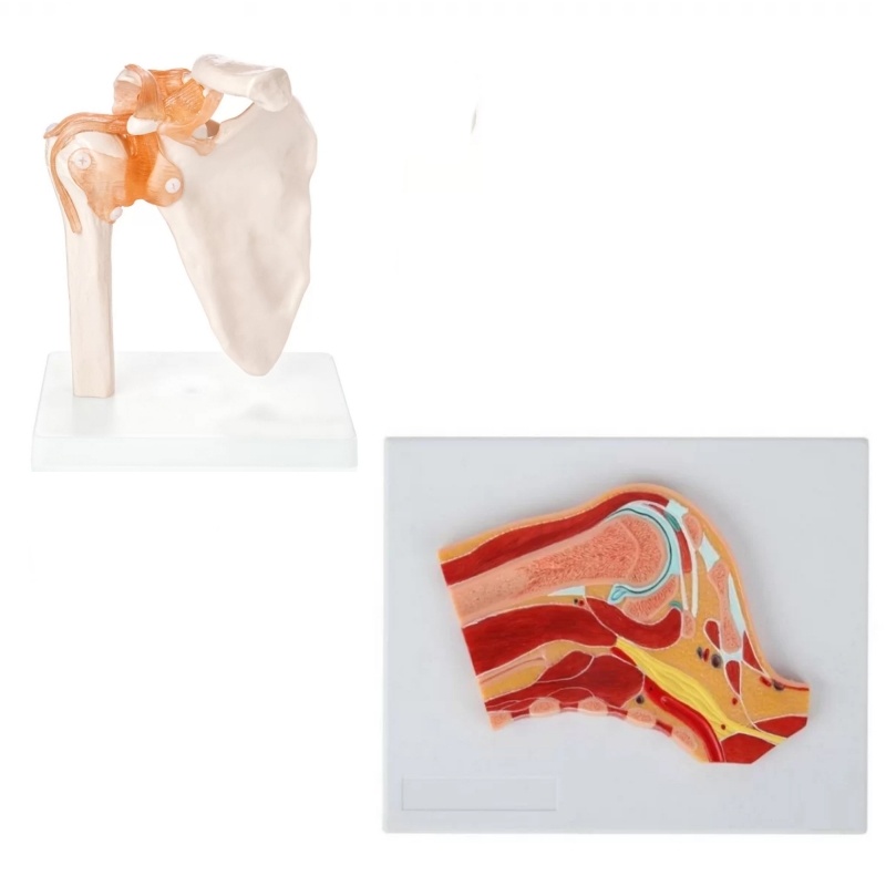
MYASKRO Shoulder Joint & Cross-Section Anatomy Model Bundle
Dual-view teaching set pairing a life-size shoulder (scapula–clavicle–proximal humerus) with an enlarged cross-section. Built for MBBS/BPT/BOT teaching, orthopedics OSCE/OSPE, and skill-lab demos.
Key Benefits
Two perspectives, one kit: Articulated glenohumeral joint plus high-contrast cross-section for instant 3D–to–sectional correlation.
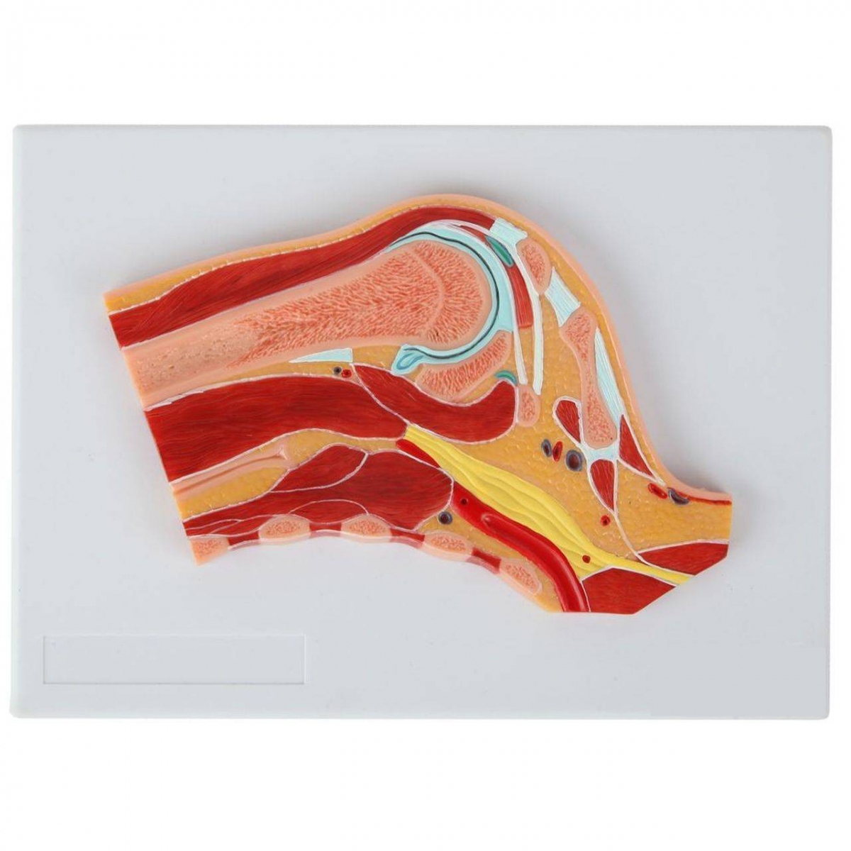
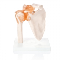
Essential anatomy shown: Glenoid fossa with labrum, articular cartilage, joint capsule, acromion/coracoid/clavicle, long-head biceps tendon, and rotator-cuff tendons (SSP/ISP/TM/SSc).
Ligamentous stability: Coracoacromial, coracoclavicular, acromioclavicular and glenohumeral ligaments highlighted for explaining stability and dislocation patterns.
Clinical correlation: Map impingement/subacromial bursitis, SLAP/Bankart lesions, rotator-cuff tears, AC joint strain; discuss safe landmarking for injections (education only).
Classroom ready: Durable, wipe-clean surfaces; colour-coded layers; stable base for benches, demos and exams.
Teach the shoulder comprehensively. A life-size joint demonstrates motion and stability while the enlarged section reveals labrum, cuff, capsule and bursae—ideal for lectures and OSCEs.
This MYASKRO bundle integrates osteology with soft-tissue planes so learners can connect palpable landmarks to sectional anatomy and imaging. The joint model clarifies humeral head–glenoid articulation, capsuloligamentous restraints and scapular relations (acromion, coracoid, clavicle). The cross-section details the rotator cuff, subacromial space/bursa, labrum and neurovascular corridors (axillary nerve/PCHA region), supporting discussions on impingement mechanisms, instability (anterior/inferior), SLAP/Bankart lesions and rehabilitation principles. Rugged materials tolerate daily handling and routine disinfection in busy skill labs.
Teaching Objectives & Skills Practised
Identify bony landmarks and rotator-cuff musculotendinous anatomy.
Explain capsuloligamentous stability and common dislocation directions.
Correlate sectional planes with X-ray/MRI and clinical tests (Neer, Hawkins, Jobe).
Demonstrate movement arcs: abduction, external/internal rotation; scapulohumeral rhythm.
Structure OSCE/OSPE shoulder examination stations using accurate models.
What’s Included
1 × Life-size shoulder joint on stand (scapula, clavicle, proximal humerus; capsule/ligaments highlighted).
1 × Enlarged colour cross-section of shoulder region on display board.