


₹ 5,999 ₹ 8,965

₹ 1,599 ₹ 4,537
")
₹ 1,699 ₹ 4,561

₹ 3,999 ₹ 7,315

₹ 1,899 ₹ 2,241
 Dissectible Into 3-Parts - Myaskro")
₹ 2,599 ₹ 7,782

₹ 5,799 ₹ 15,157

₹ 8,499 ₹ 22,445

₹ 4,199 ₹ 10,555
 5.8 Feet Tall 96% Anatomical Accuracy Premium Quality | MYASKRO")
₹ 14,699 ₹ 33,039

₹ 11,799 ₹ 22,379

₹ 7,999 ₹ 14,750

₹ 8,199 ₹ 22,379

₹ 20,599 ₹ 28,965

₹ 5,999 ₹ 8,945

₹ 11,799 ₹ 18,945
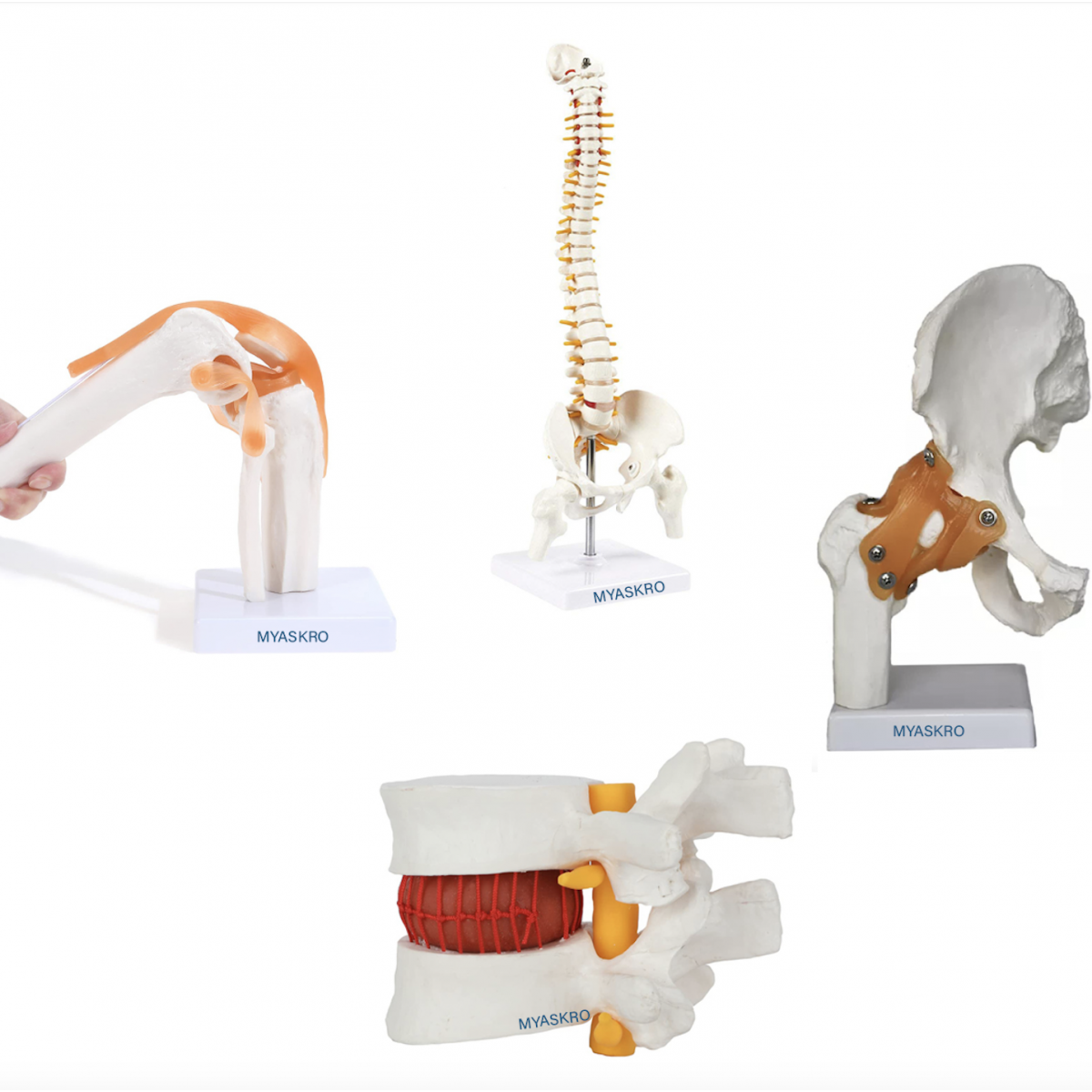
When you're teaching human anatomy or guiding patient care, clarity matters. The MYASKRO Human Joints Anatomical Models – Set of 4 is a premium, comprehensive kit that includes life-size replicas of the knee joint, spinal column, hip joint, and lumbar disc herniation. Each model is crafted with clinical accuracy and mounted for convenient tabletop use.
This set is ideal for medical colleges, physiotherapy institutions, orthopedic clinics, rehabilitation centers, and surgical demo labs. It provides everything needed to demonstrate structure, movement, injury mechanisms, and postural education—all in one cohesive, portable bundle.
Included in the set:
All models are made from non-toxic, medical-grade PVC, designed for high-traffic use and repeated demonstrations. Each sits firmly on its own display base, allowing for individual handling or group viewing during lectures and consultations.
Searchers looking for “orthopedic joint models,” “set of anatomical teaching tools,” “PVC spine and hip replica,” or “medical education joint models in India” will find this kit offers unmatched value and anatomical accuracy.
“We use this combo set during clinical rounds for orthopedic interns—it’s made a huge difference in how quickly they grasp biomechanics and injury diagnosis,” says Dr. Iqbal Khan, Professor of Orthopedics, GMC Nagpur.
For anyone seeking to elevate their anatomical teaching, improve patient communication, or train the next generation of clinicians—this set is the ultimate visual toolkit.
This MYASKRO set brings together four of the most critical joint models in clinical and anatomical education—allowing educators and professionals to teach structure, pathology, and movement with hands-on precision. Each model is designed to function independently or in tandem with others, giving you complete flexibility in lesson planning, diagnosis, and demonstration.
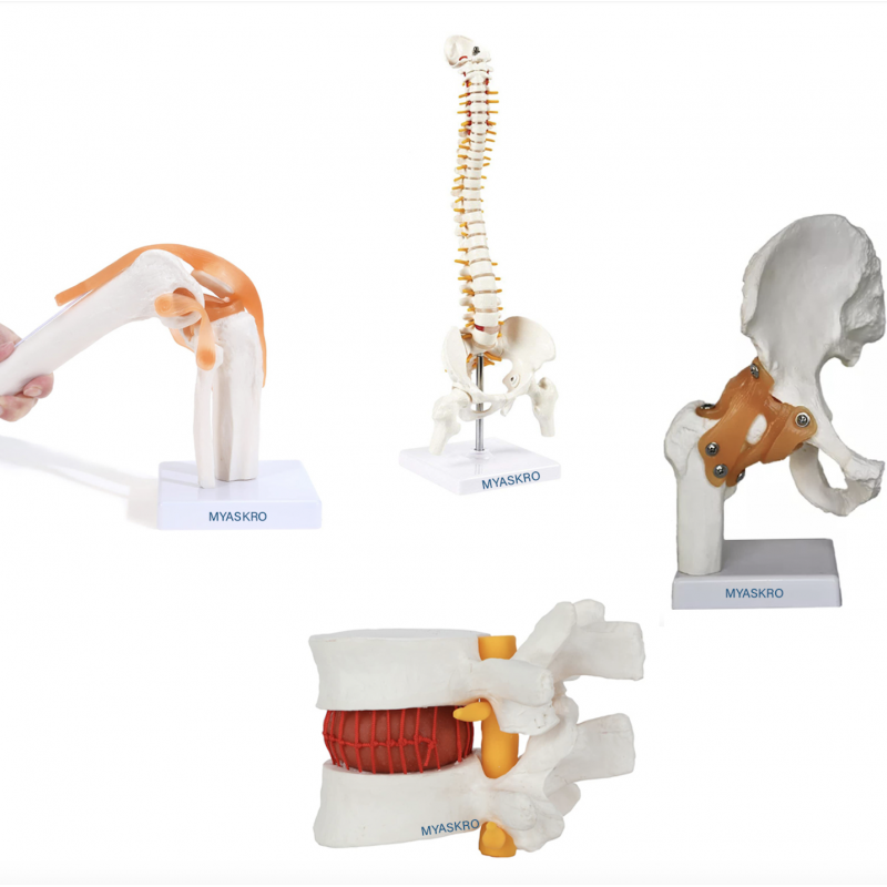
1. Knee Joint Model: With detailed rendering of the femur, tibia, fibula, and patella, this model includes:
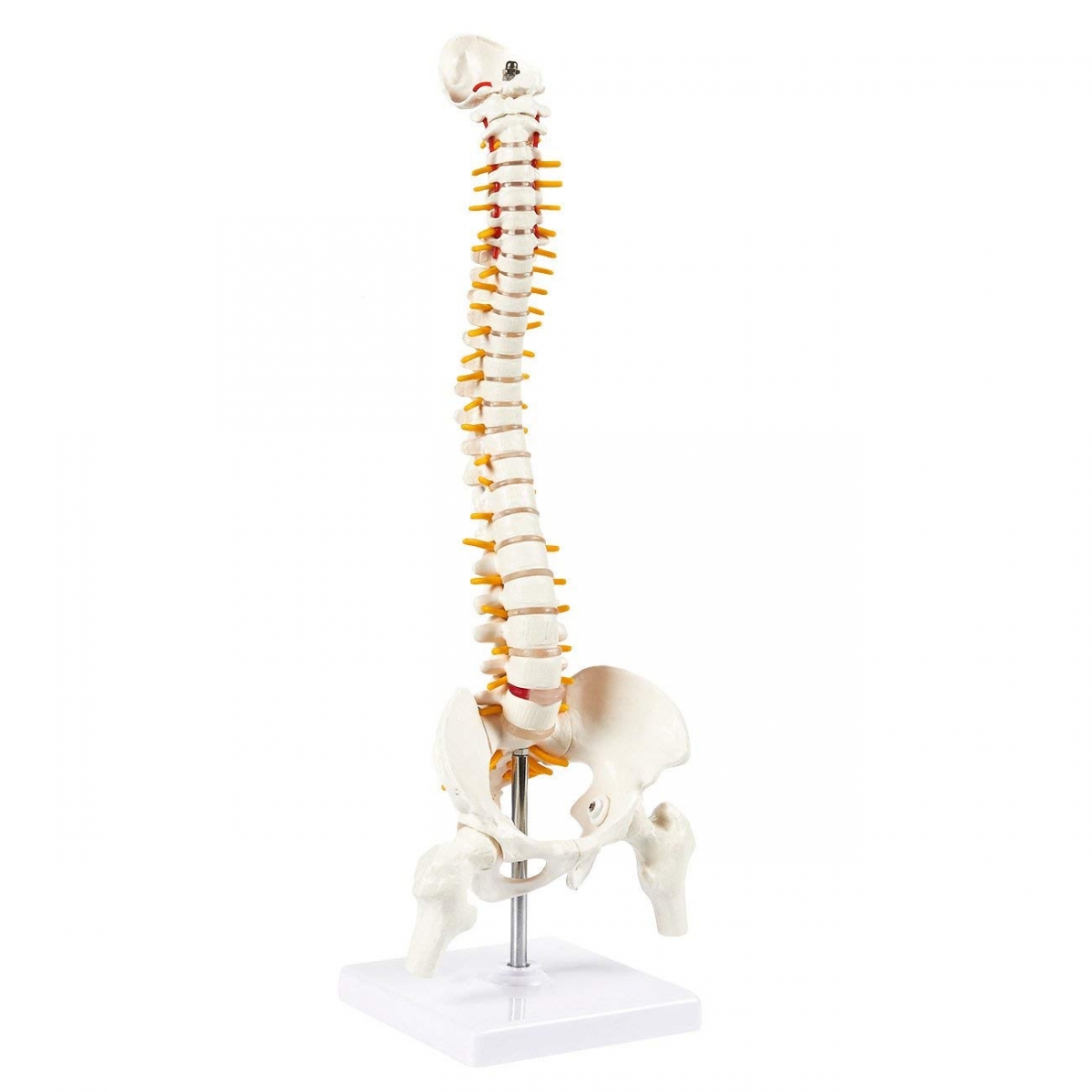
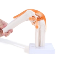
2. Spine with Pelvis Model: Cervical to sacral spine, featuring:
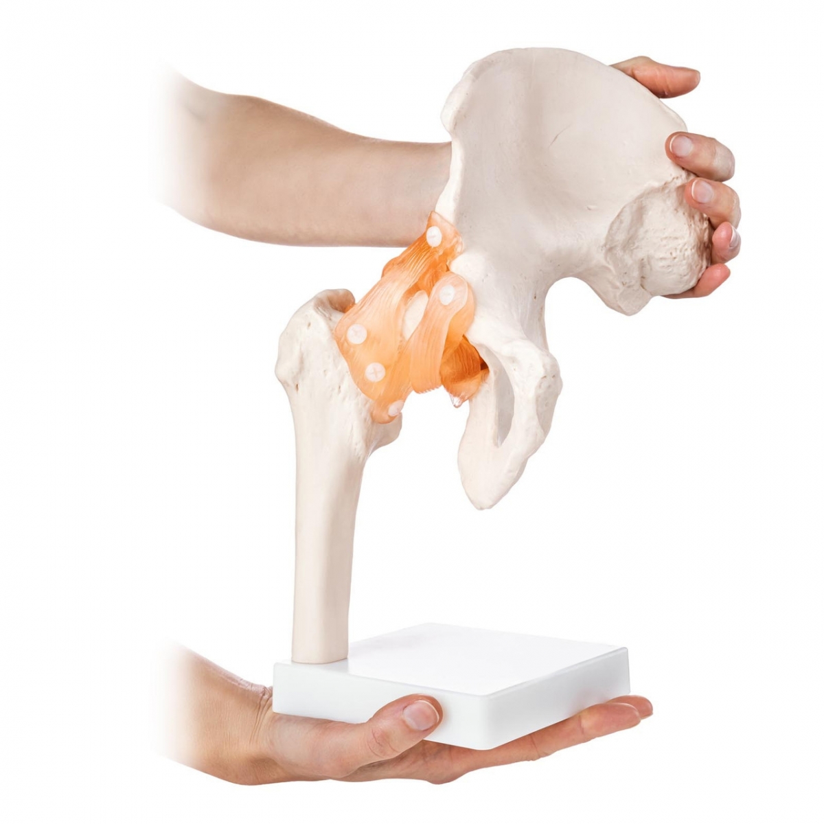
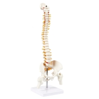
3. Hip Joint Model with Ligaments: Shows the relationship between femoral head and pelvic acetabulum, featuring:
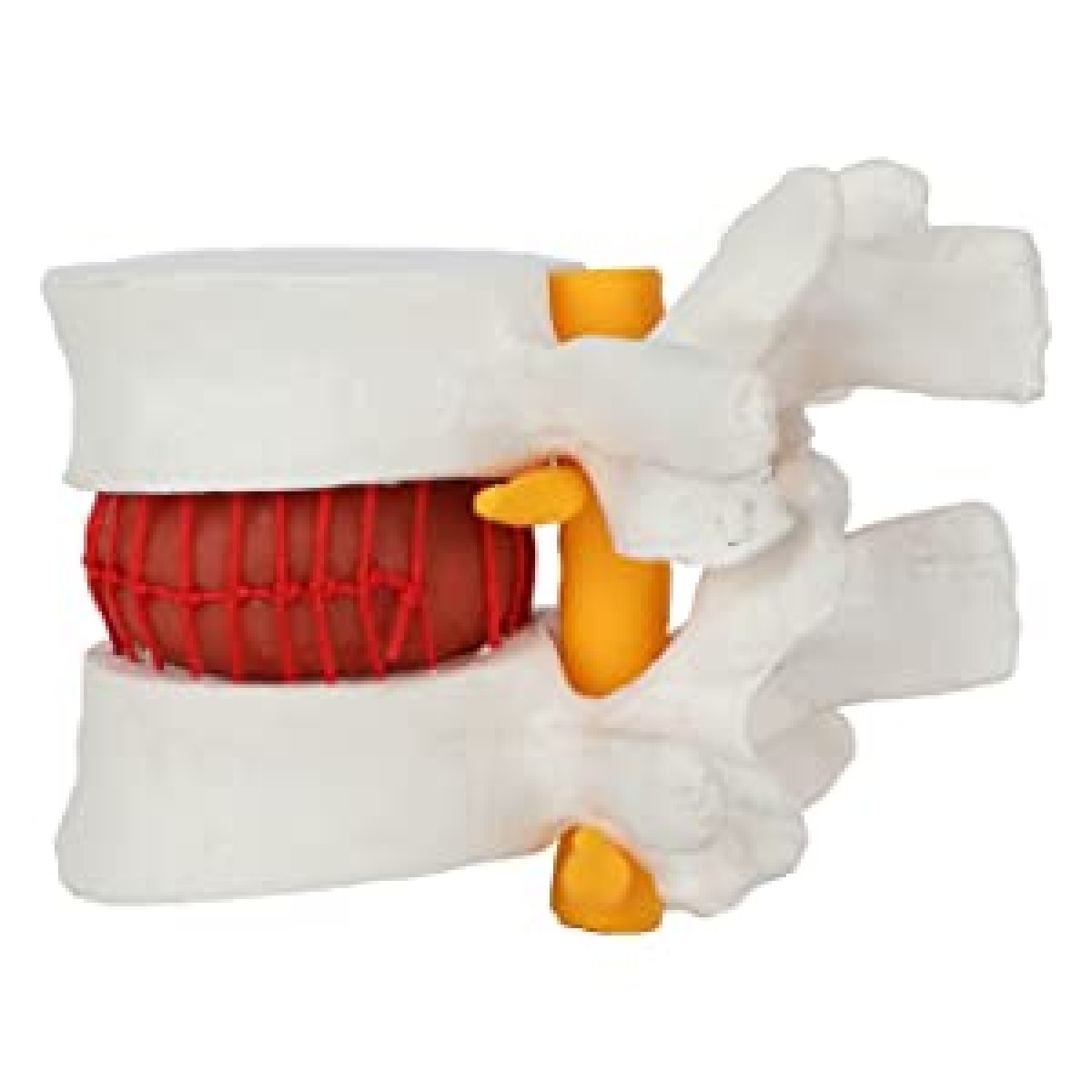
4. Lumbar Disc Herniation Model: A focused model of the L4-L5 segment, clearly illustrating:
All models are crafted using medical-grade PVC and durable soft-tissue polymer to allow both realism and resilience. They stand independently on clean, labeled bases, making them suitable for rotating use in busy clinical or classroom settings.
“We’ve integrated this set into our BPT program at Jamia Hamdard, Delhi, and students have dramatically improved their understanding of joint pathology,” shares Prof. Aashima Rathi, PT Faculty.
Each model in this set is a powerful teaching asset—but together, they form a complete visual system for musculoskeletal education and joint-focused clinical discussions.
In a world where anatomy education and orthopedic care demand clarity, the MYASKRO Set of 4 Human Joint Models delivers the tools needed to educate with confidence. Each model in this collection brings a vital region of the body into focus, from the stabilizing ligaments of the knee to the intricate spinal column and the deep-set mechanics of the hip and lumbar vertebrae.
This set empowers professors, clinicians, therapists, and students to bridge the gap between textbook theory and real-life function. You can physically demonstrate:
Each model is crafted for daily handling and demonstration. They're light enough to move from classroom to clinic, yet sturdy enough for high-use environments. The joints retain their anatomical form even after repeated manipulation, ensuring your investment remains dependable over time.
This kit also aligns perfectly with high-intent keywords such as “orthopedic joint model combo,” “life-size PVC anatomy models,” “spine and joint demonstration set,” and “physiotherapy joint teaching tools.” It fulfills every need those searches suggest—whether you're instructing MBBS students or guiding a patient through pre-surgical orientation.
“This 4-model set has become an integral part of our orthopedic OPD. Whether it's explaining disc herniation or knee instability, these visuals save us time and improve patient understanding,” says Dr. Neha Tyagi, Consultant Ortho, Fortis Hospital, Noida.
Some tools assist. Others transform. This MYASKRO joint model set falls firmly in the latter category. With it, every session becomes more interactive, every explanation more precise, and every learner—whether student or patient—walks away with a clearer picture of the human body in motion.
Total Reviews (0)