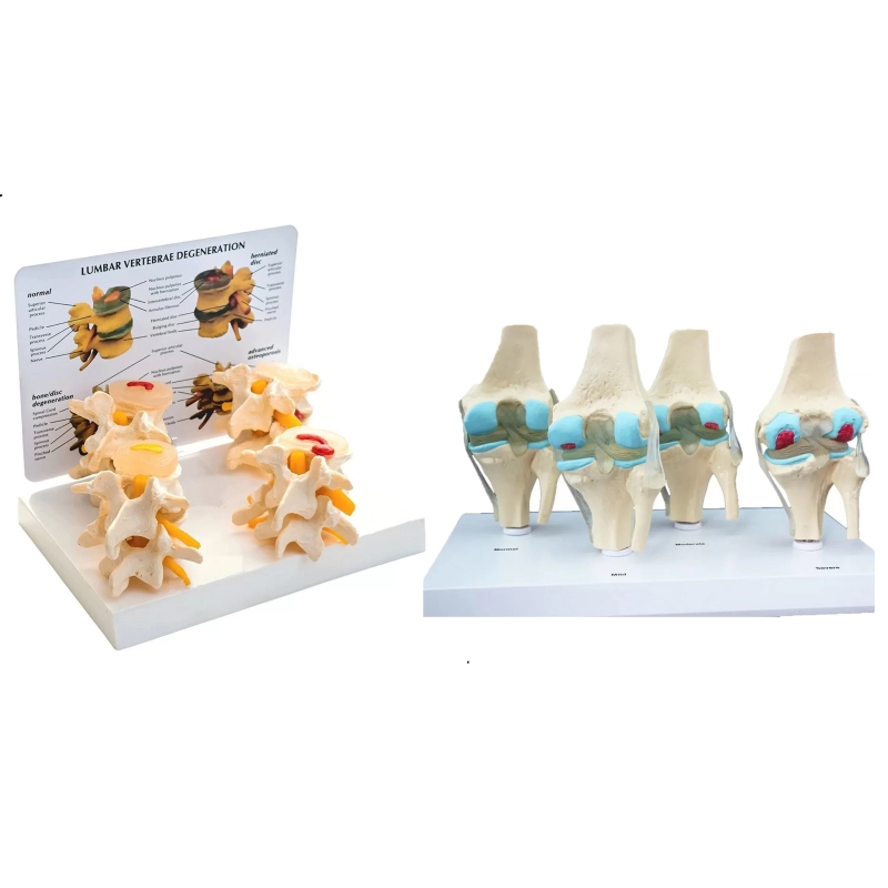


4-Stage Lumbar Vertebral Degeneration Model + 4-Stage Knee Degeneration Model
Components
Lumbar vertebral degeneration (4 stages): Lumbar motion segments with intervertebral discs and exiting spinal nerve roots. Stages illustrate normal disc, annular bulge (protrusion) with foraminal encroachment, disc extrusion (herniation) with root compression, and advanced degenerative disc disease with disc-height loss, osteophytes, and canal/foraminal narrowing.
Knee degeneration (4 stages): Distal femur–proximal tibia–patella showing progression from normal articular cartilage to cartilage thinning/fibrillation, meniscal degeneration/tear, joint-space narrowing with marginal osteophytes and subchondral sclerosis (osteoarthritis cascade).
Learning objectives
Correlate structural change with symptoms/signs: radiculopathy from foraminal stenosis; mechanical knee pain, crepitus, reduced ROM in OA.
Explain pathophysiology: disc dehydration → annular fissure → protrusion/extrusion; inflammatory–mechanical cycle of knee OA.
Demonstrate imaging correlations (MRI/weight-bearing radiographs) and discuss indications for conservative therapy, injections, or surgical referral (teaching context).
Specifications
Teaching scale models in rigid PVC on labeled bases; stable display.
Cleaning: mild detergent or 70% alcohol wipe; avoid solvents/heat.
Intended use: orthopaedics, physiatry, physiotherapy, pain clinic, UG/PG anatomy, OSCE/skill-lab, and patient education.
Total Reviews (0)