


₹ 3,999 ₹ 7,315

₹ 20,999 ₹ 27,655

₹ 12,599 ₹ 25,836

₹ 7,999 ₹ 14,750

₹ 11,799 ₹ 22,379
 | Clinical-Grade Wall Charts - Myaskro")
₹ 6,399 ₹ 8,006

₹ 17,499 ₹ 32,975

₹ 22,299 ₹ 57,779
 With Special Lamination")
₹ 1,599 ₹ 3,865
 With Special Lamination")
₹ 1,599 ₹ 3,865
 With Hard Backing, Aluminium Frame & Hanging Hooks")
₹ 2,800 ₹ 4,561

₹ 1,599 ₹ 4,955
 With Ligaments")
₹ 1,699 ₹ 4,955

₹ 8,199 ₹ 22,379

₹ 5,999 ₹ 8,945
")
₹ 6,599 ₹ 8,945
")
₹ 6,399 ₹ 8,675

₹ 20,599 ₹ 28,965
")
₹ 9,499 ₹ 17,845

₹ 5,999 ₹ 8,945

₹ 11,799 ₹ 18,945
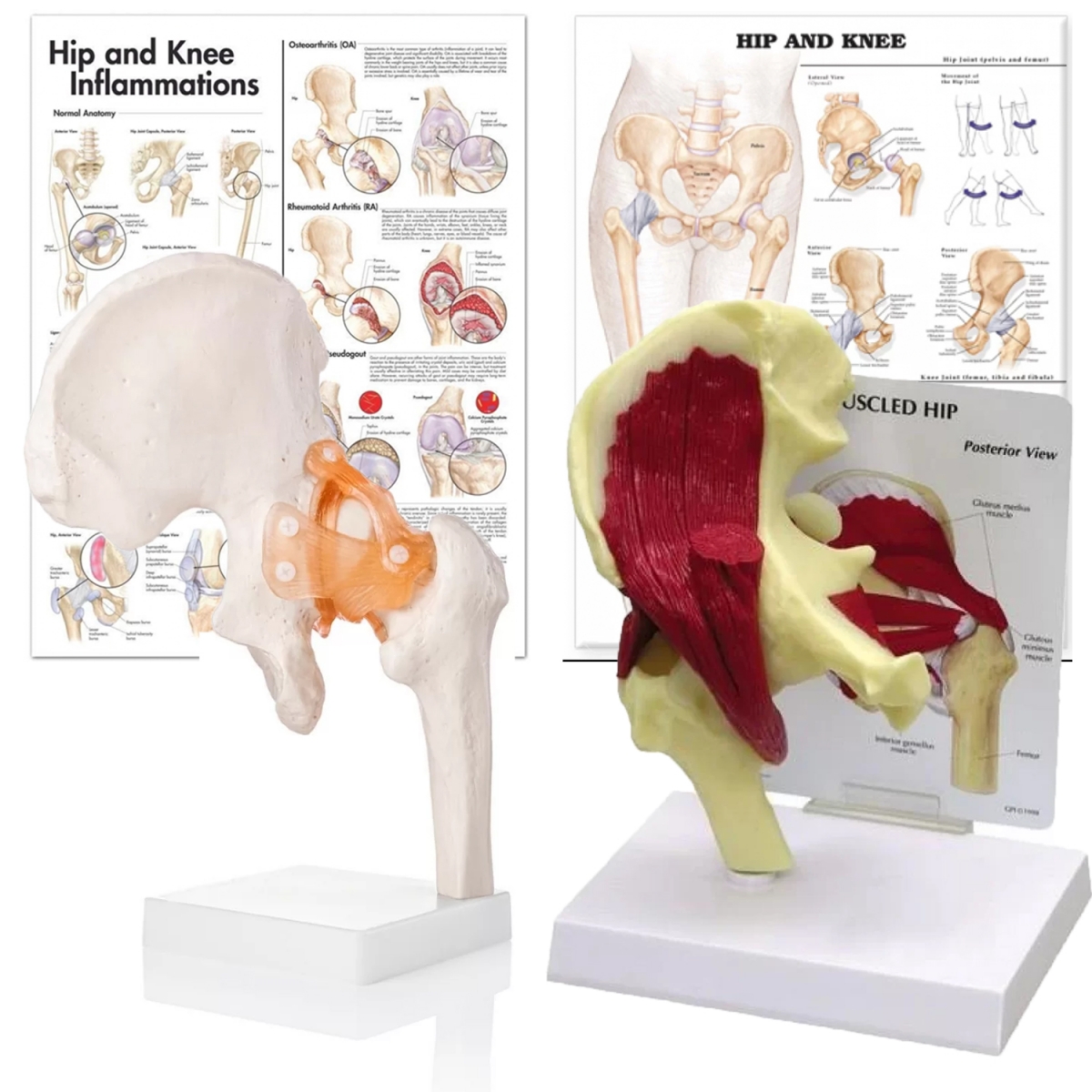
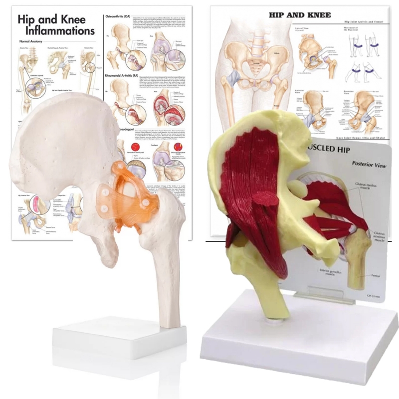
Components
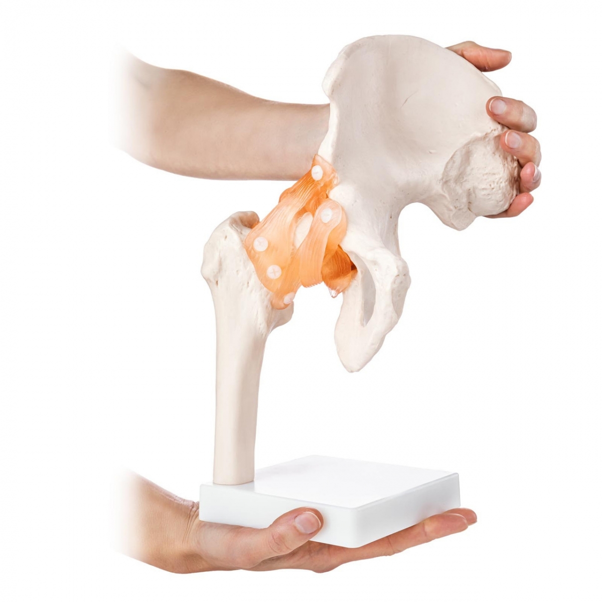
Hip joint with ligaments: Pelvis (ilium–ischium–pubis) with proximal femur; capsule with iliofemoral, pubofemoral, ischiofemoral ligaments; modeled transverse acetabular ligament; articulated head–neck for demonstration of physiologic ROM.
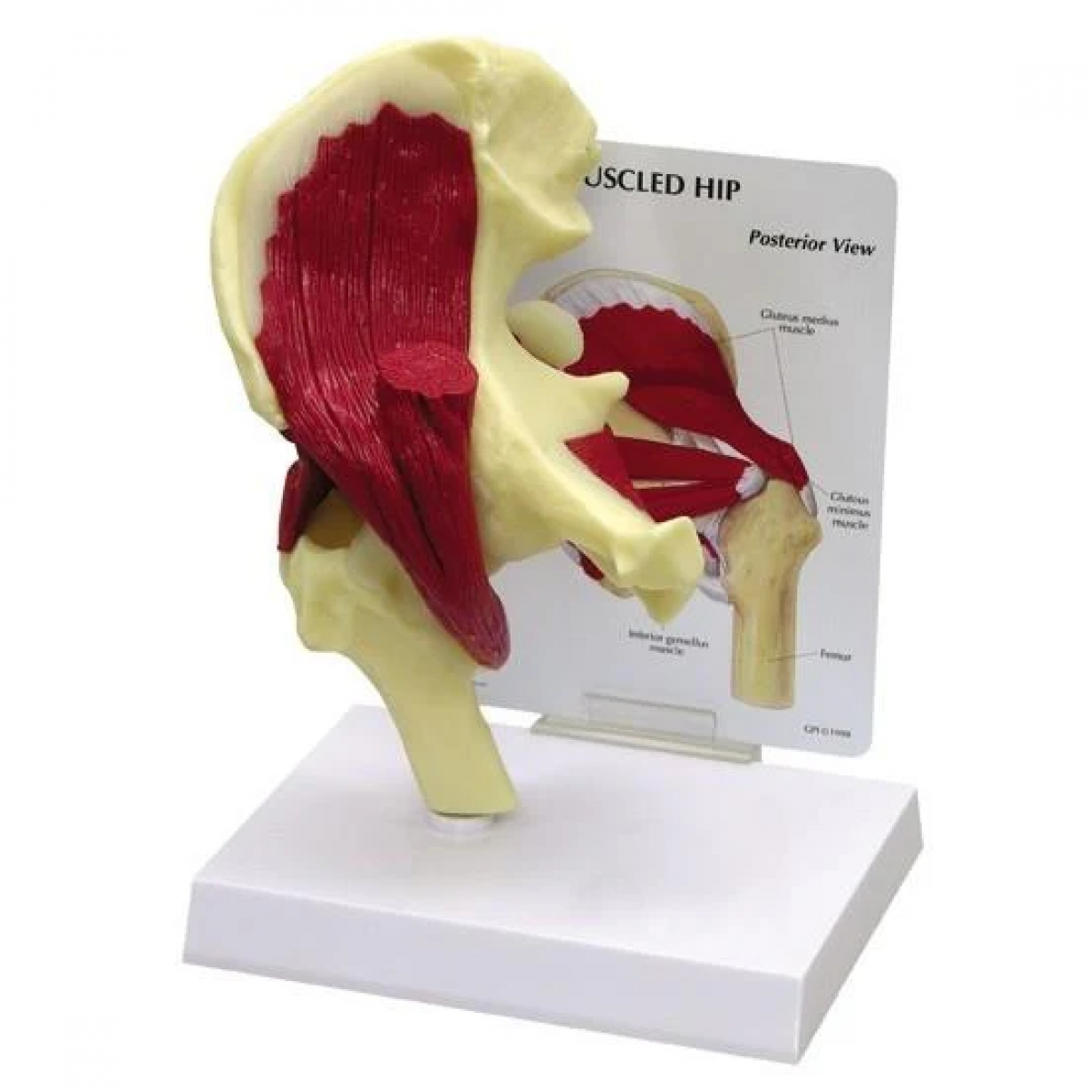
Hip joint with muscles: Superficial myology of gluteus maximus, medius, minimus, tensor fasciae latae, and deep external rotators (piriformis, obturator internus/externus, gemelli, quadratus femoris); visible greater/lesser trochanter and muscle insertions.
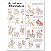
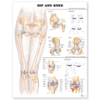
Charts (52 × 70 cm, laminated with rollers):
Hip & Knee (osteology, axes of movement)
Hip & Knee Inflammations (OA, RA, bursitis, tendinopathy; schematic pathology)
Learning objectives
Identify acetabulum, femoral head–neck, greater/lesser trochanters, ligament attachments, and capsular fiber orientation.
Explain roles of iliofemoral (Y) ligament in resisting extension/external rotation; pubofemoral in limiting abduction/extension; ischiofemoral in controlling internal rotation.
Correlate abductor mechanism (gluteus medius/minimus, TFL) with pelvic stability and Trendelenburg gait.
Discuss common pathology: hip OA, RA synovitis, trochanteric bursitis, tendinopathy of abductors, and external rotator syndrome.
Set up OSCE tasks: landmark identification, ligament function, abductor testing, safe ROM demonstration.
Specifications
Scale: life-size; material: Medical Grade PVC with color-coded structures; individual base stands.
Charts: heavy-gauge lamination (dry-wipe), top–bottom rollers.
Care: wipe with mild detergent or 70% alcohol; avoid solvents/heat.
Intended use: instructional aid for orthopaedics, physiotherapy, sports medicine, UG/PG anatomy.
Total Reviews (0)