


₹ 1,599 ₹ 4,537

₹ 5,999 ₹ 8,965

₹ 5,999 ₹ 7,645

₹ 7,999 ₹ 22,379

₹ 7,999 ₹ 14,750

₹ 11,799 ₹ 22,379
 With Special Lamination")
₹ 1,599 ₹ 3,865
 With Special Lamination")
₹ 1,599 ₹ 3,865
 With Special Lamination")
₹ 1,599 ₹ 3,865
 With Hard Backing, Aluminium Frame & Hanging Hooks")
₹ 2,800 ₹ 4,561
 With Hard Backing, Aluminium Frame & Hanging Hooks")
₹ 2,800 ₹ 4,561

₹ 1,499 ₹ 4,955

₹ 1,699 ₹ 4,955
 With Ligaments")
₹ 1,699 ₹ 4,955
")
₹ 1,699 ₹ 4,561

₹ 6,399 ₹ 15,326
")
₹ 3,199 ₹ 6,495

₹ 11,499 ₹ 12,865
")
₹ 6,399 ₹ 8,675

₹ 20,599 ₹ 28,965
")
₹ 4,799 ₹ 6,245
 | Myaskro")
₹ 3,199 ₹ 4,865

₹ 11,799 ₹ 18,945
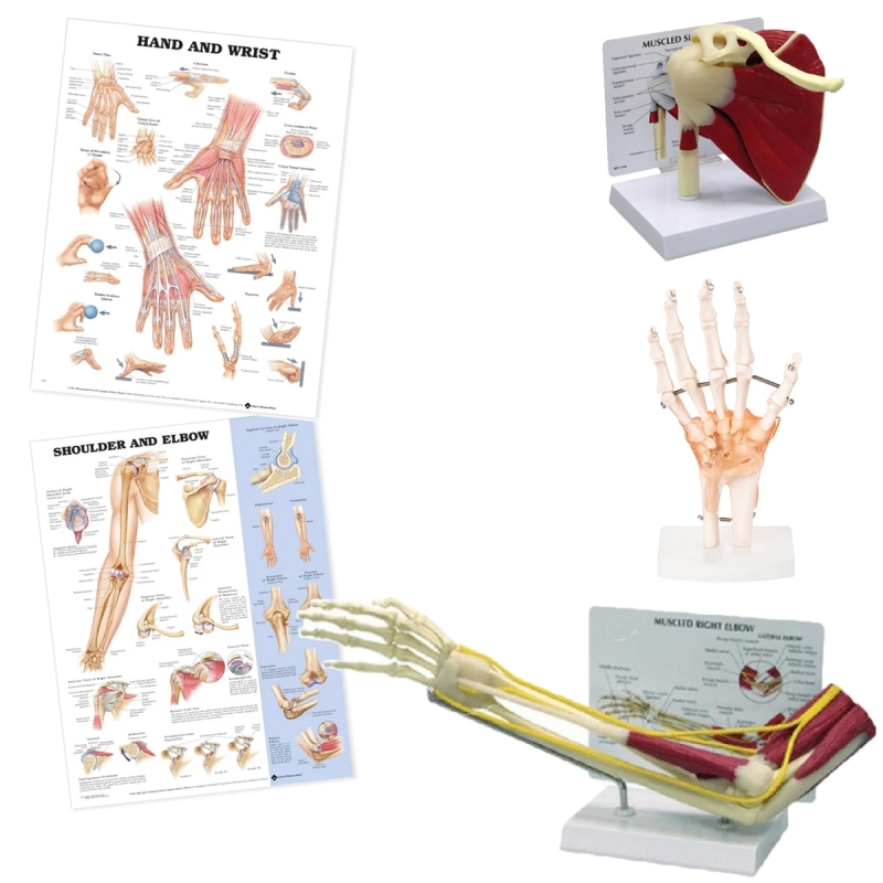
Components
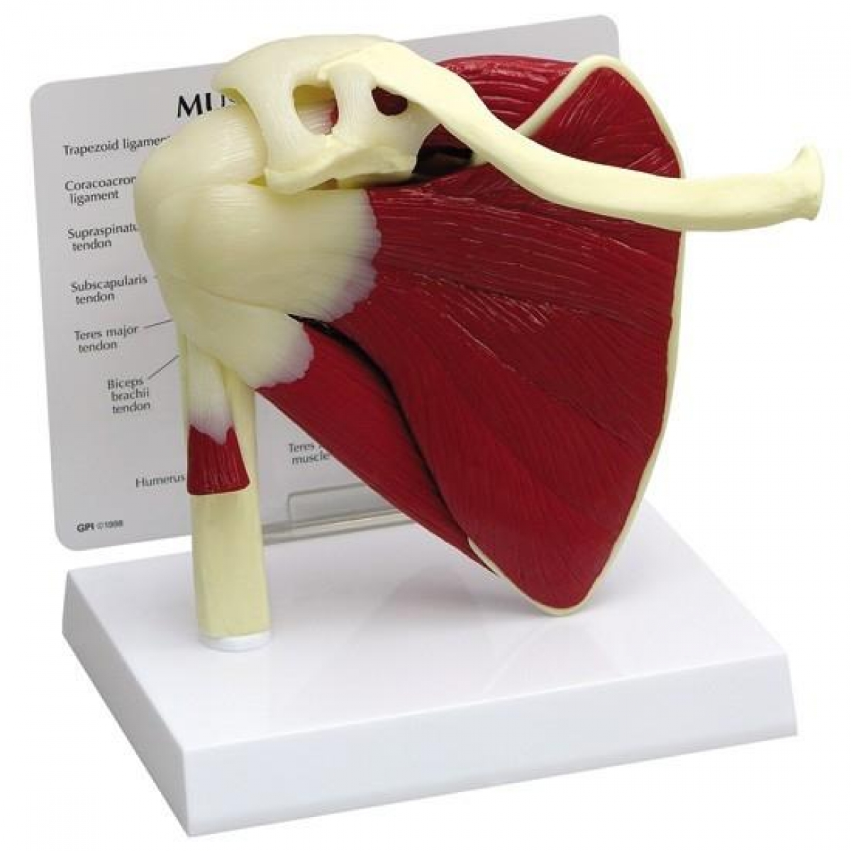
Muscular shoulder joint model: Scapula–clavicle–proximal humerus with rotator-cuff muscles (supraspinatus, infraspinatus, teres minor, subscapularis) and deltoid; capsule/space orientation for impingement discussion.
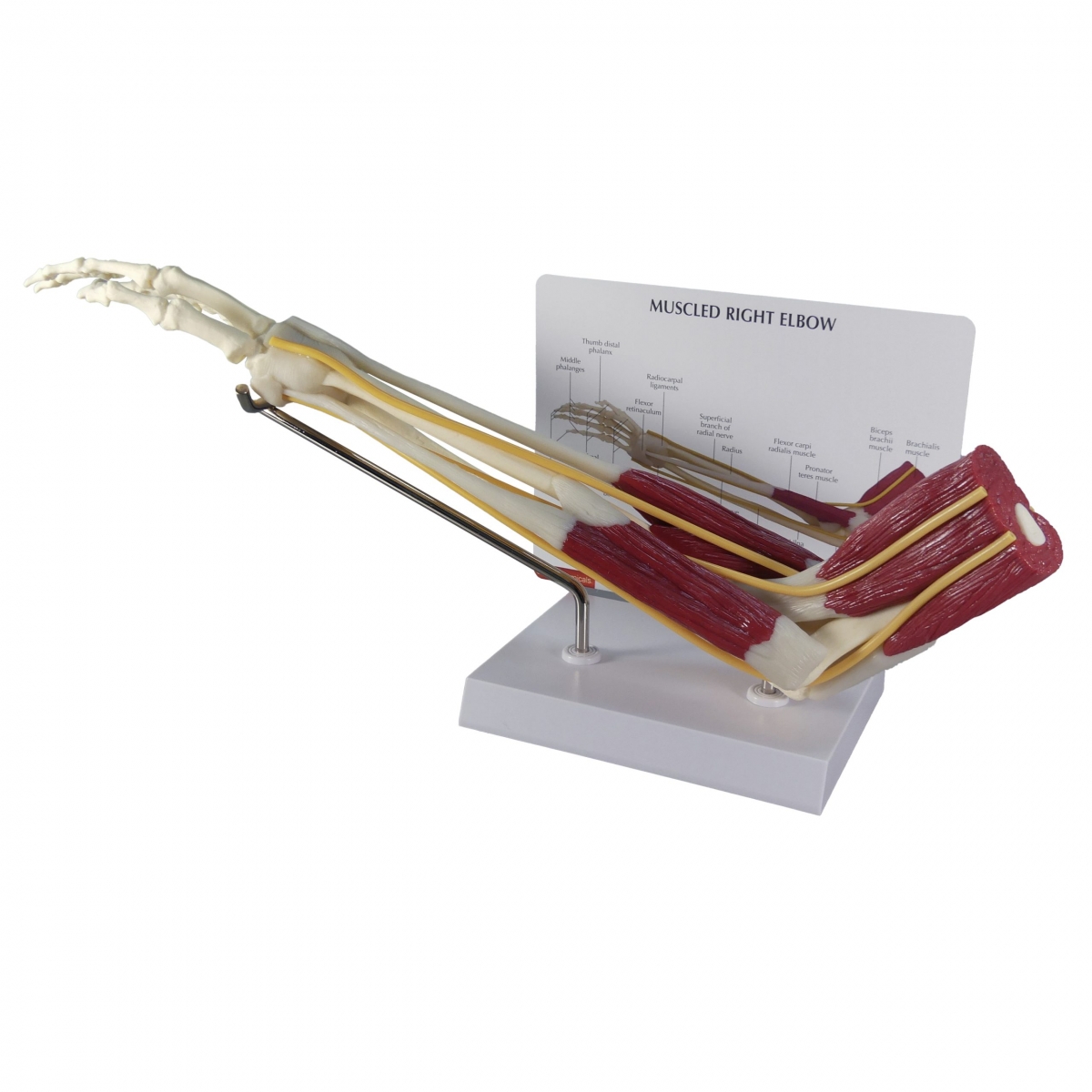
Muscular elbow joint model: Humerus–radius–ulna with biceps, brachialis, triceps, common flexor/extensor tendons, major nerves (median, ulnar, radial) routed to the forearm.
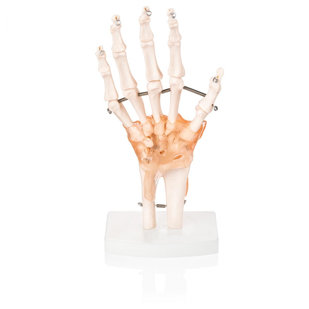
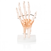
Hand joint with ligaments: Carpus–metacarpus–phalanges showing collateral ligaments, volar plates, flexor/extensor retinacula, and carpal tunnel limits.
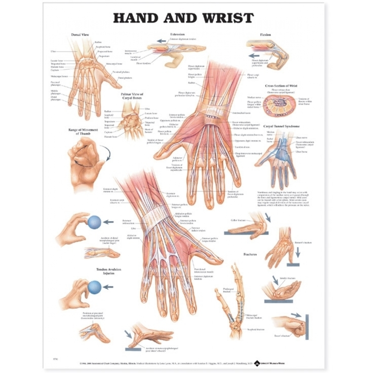
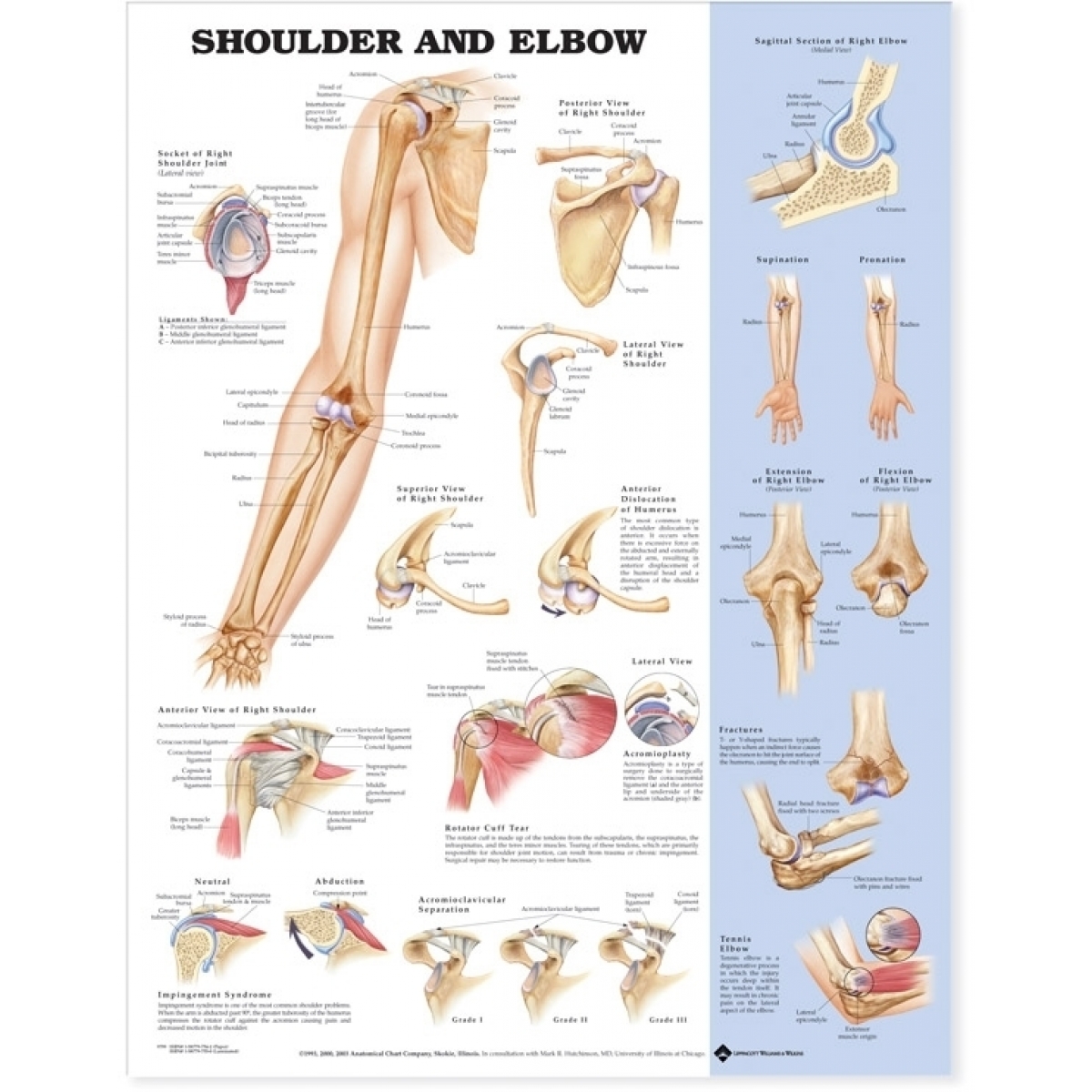
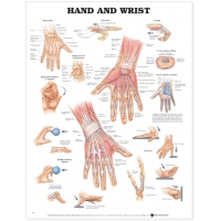
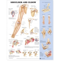
Charts (52 × 70 cm, laminated with rollers): Hand & Wrist Anatomy and Shoulder & Elbow (osteology, myology, movements).
Patient-education objectives
Explain shoulder kinematics (glenohumeral, scapulothoracic rhythm) and pathology: rotator-cuff tendinopathy/tear, subacromial impingement, frozen shoulder.
Demonstrate elbow stability (UCL/RCL/annular ligaments), common conditions: lateral/medial epicondylitis, olecranon bursitis, radial head injury.
Visualize wrist/hand stabilizers and carpal alignment; counsel on carpal tunnel syndrome, De Quervain’s, UCL injury (skier’s thumb), trigger finger, OA/RA changes.
Use models to show safe ROM, nerve entrapment sites, and post-op/rehab precautions.
Specifications
Life-size, Medical Grade PVC models on labeled bases; high-contrast, hand-painted detail.
Charts: heavy-gauge, dry-wipe lamination with top–bottom rollers.
Cleaning: mild detergent or 70% alcohol; avoid solvents/heat.
Intended users
Outpatient clinics, physiotherapy/OT, orthopaedics, sports medicine, and medical colleges for bedside counselling and OSCE teaching.
Total Reviews (0)