

₹ 3,999 ₹ 7,315

₹ 1,599 ₹ 4,955

₹ 5,299 ₹ 8,011
 With Special Lamination")
₹ 1,599 ₹ 3,865

₹ 17,499 ₹ 32,975

₹ 33,599 ₹ 57,779

₹ 8,699 ₹ 22,379

₹ 12,599 ₹ 25,836

₹ 20,999 ₹ 27,655

₹ 20,599 ₹ 28,965

₹ 11,799 ₹ 18,945
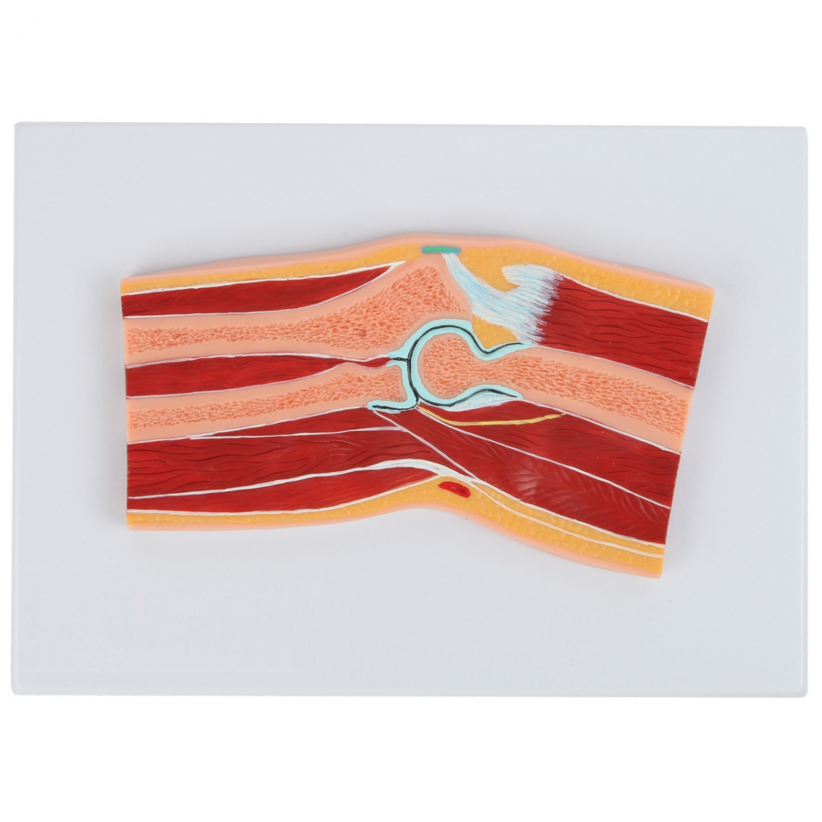
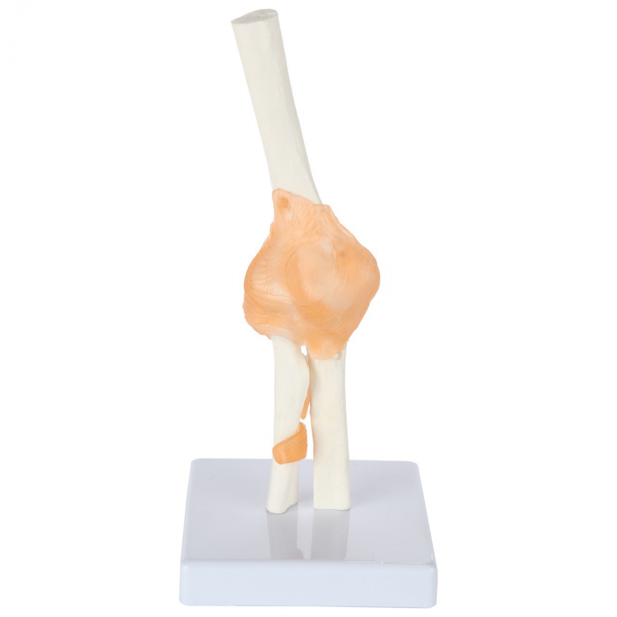
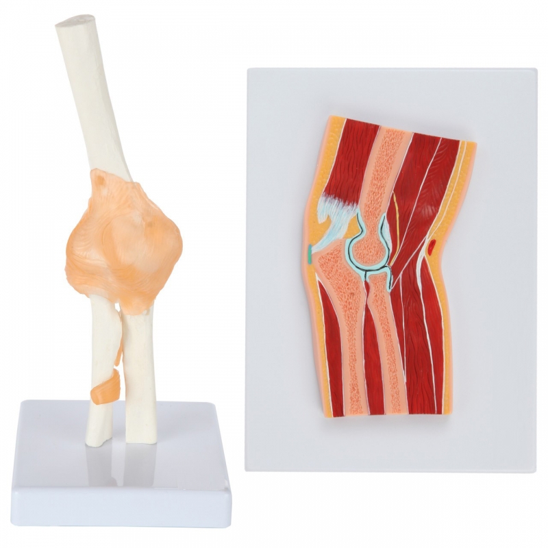
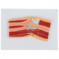
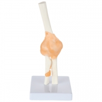
A compact, clinic-ready set that makes explaining elbow structure and injuries fast and visual. The articulated elbow demonstrates motion and ligament relationships, while the colorful cross-section plaque shows the layered anatomy at a glance.
Functional Elbow Joint Model (life-size, on base) – Humerus, radius, and ulna with flexible ligaments to demonstrate flexion/extension and pronation/supination.
Elbow Cross-Section Relief Model (enlarged, on board) – Raised, color-coded section showing skin, subcutaneous tissue, muscles, tendons, joint capsule, hyaline cartilage, nerves, and vessels.
Hands-on motion: Show common issues such as sprains, epicondylitis, bursitis, and radius/ulna alignment during rotation.
Layered understanding: Cross-section clarifies where tendons, nerves, and bursae sit relative to the joint.
Durable & easy to clean: Cast in high-quality PVC with wipe-clean surfaces.
Ready to display: Each model mounted on a sturdy base/board; ideal for desks or wall display (cross-section).
Patient-friendly colors & labels: Clear, non-intimidating visualization for quick education.
Orthopedic & sports clinics, physiotherapy, chiropractic, athletic training rooms, classrooms, and OSCE prep.
Material: Medical-grade PVC, metal support rod (joint), printed relief board (cross-section)
Scale: Joint model life-size; cross-section enlarged for clarity
Care: Wipe with mild soap solution; avoid solvents