

Components
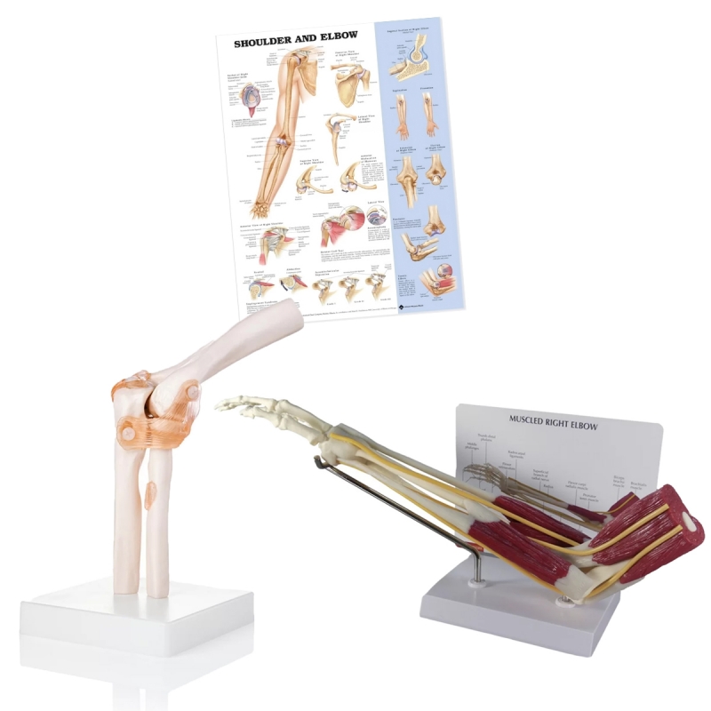
Elbow with ligaments: Humerus–ulna–radius with joint capsule, ulnar collateral ligament (anterior/posterior/transverse bands), radial collateral ligament, annular ligament stabilising the proximal radioulnar joint; demonstration of valgus/varus constraints and pronation–supination.
Elbow with muscles: Anterior and posterior compartments with biceps brachii (radial tuberosity), brachialis (ulnar tuberosity), triceps (olecranon), brachioradialis, and common flexor/extensor tendons at the epicondyles; visible median, radial, ulnar nerves and forearm tendons on an angled teaching stand.
Chart (52 × 70 cm, laminated with rollers): Shoulder & Elbow—osteology, axes of movement, myology, and common lesions.
Learning objectives
Identify bony landmarks: capitulum, trochlea, coronoid/olecranon fossae, radial head/neck, ulnar/radial tuberosities.
Explain stability: UCL for valgus restraint; RCL/annular for varus and posterolateral rotatory stability; capsule in end-range extension.
Correlate pathology: lateral/medial epicondylitis, UCL injury (throwers), nursemaid’s elbow, olecranon bursitis, radial head fracture.
Demonstrate mechanics: flexion–extension at humeroulnar/humeroradial joints; pronation–supination at PRUJ; carrying angle assessment.
OSCE stations: ligament identification, resisted tests (Cozen/Mill; golfer’s), landmark palpation, nerve course orientation.
Specifications
Life-size medical grade PVC models on stable bases; numbered structures with legend card.
Chart: heavy-gauge lamination (dry-wipe), top–bottom rollers.
Care: wipe with mild detergent or 70% alcohol; avoid solvents/heat.
Use: orthopaedics, sports medicine, physiotherapy, UG/PG anatomy, and patient education.
Total Reviews (0)