


₹ 6,399 ₹ 15,039
")
₹ 6,499 ₹ 9,982

₹ 33,899 ₹ 69,579

₹ 9,599 ₹ 17,305

₹ 20,999 ₹ 27,655

₹ 7,950 ₹ 15,299

₹ 8,499 ₹ 22,445

₹ 7,999 ₹ 14,750

₹ 11,499 ₹ 12,865

₹ 20,599 ₹ 28,965

₹ 11,799 ₹ 18,945
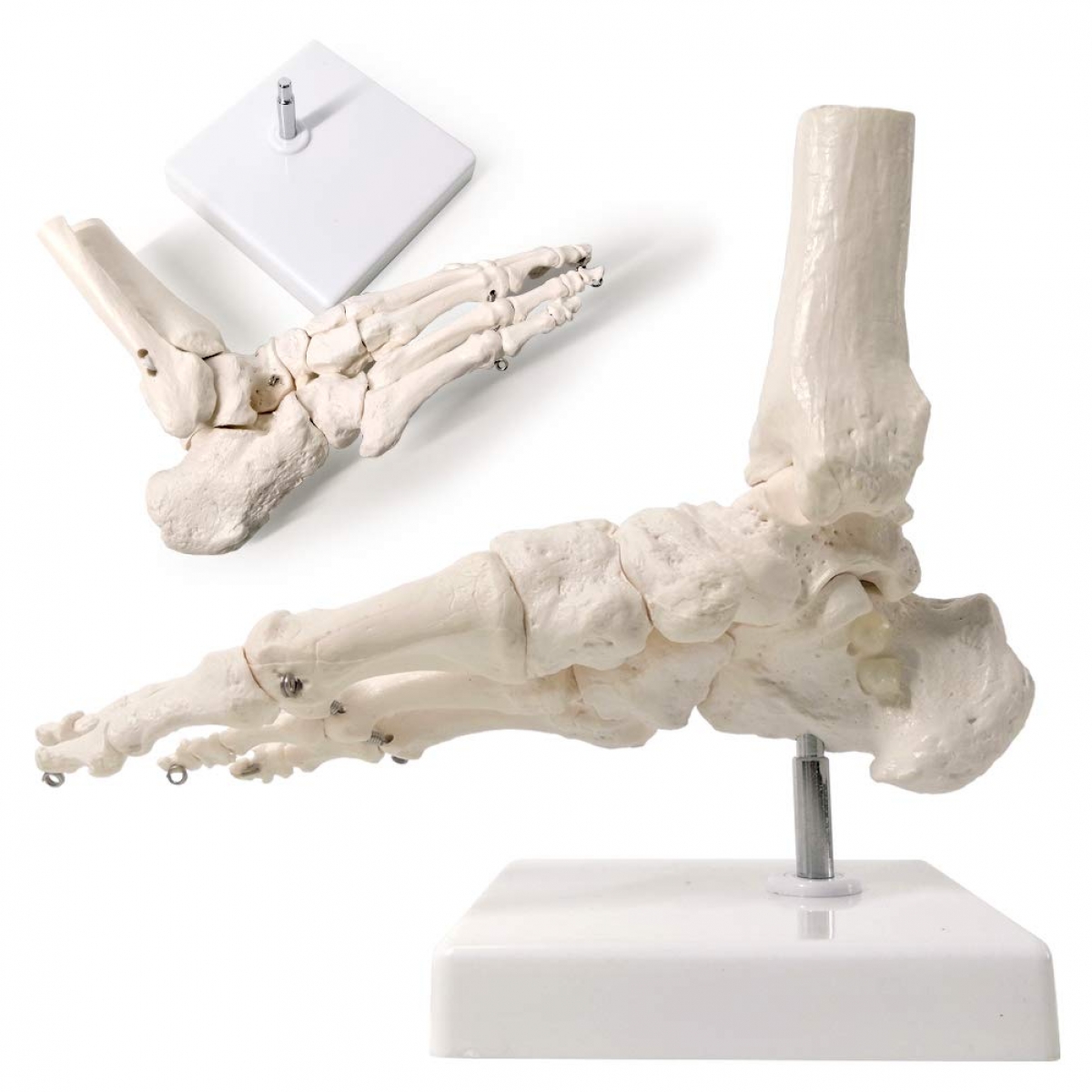
When it comes to understanding lower limb function, few structures are as complex—or as critical—as the human foot. The Life-Size Foot Joint Model offers a realistic, clinically accurate view of the skeletal anatomy that forms the foundation of posture, balance, and mobility. Built for precision and designed for practicality, this model is an indispensable tool for medical educators, physiotherapists, podiatrists, and orthopedic professionals.
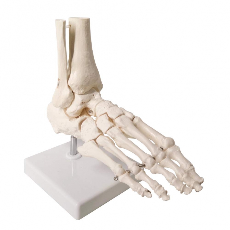
Featuring every major joint and bone of the foot—including the tibia, fibula, tarsals, metatarsals, and phalanges—this model clearly demonstrates how structural integrity affects function. Mounted on a durable base, it maintains anatomical position for easy reference and smooth rotation during lectures, exams, and patient consultations.
This model brings real-world clarity to the classroom and clinic alike. Chiropractic colleges, osteopathic programs, and sports science faculties use it to break down the mechanics of foot movement, joint stability, and weight-bearing dynamics. Clinicians use it to help patients visualize conditions like arthritis, fractures, plantar fasciitis, or bunions.
Common question: “Does it show functional joint mobility?” While the bones are fixed in anatomical position for accuracy, the visible articulation points and clear segmentation of joints make it easy to explain range of motion, joint loading, and alignment issues.
From a semantic perspective, it ranks highly for search queries such as “life-size skeletal foot model,” “foot and ankle anatomy replica,” “orthopedic joint education tool,” and “podiatry foot skeleton teaching aid.” It fits perfectly into environments where anatomical precision matters most.
“We use it during every lower limb module. Students grasp bone landmarks and joint connections much faster when they can physically examine them,” says Dr. Kashish., Lecturer in Human Anatomy, Raipur.
Compact, professional, and medically detailed, the MYASKRO Foot Joint Model doesn’t just illustrate the bones of the foot—it reinforces the role this often-overlooked region plays in every step, every jump, and every moment of upright life. A visual asset for serious anatomy learners and clinicians alike.
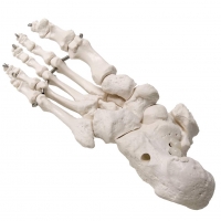
Every bone tells a story—and in the human foot, there are 26 bones working in concert to support motion, absorb impact, and maintain posture. The Life-Size Foot Joint Model captures this intricate skeletal architecture with remarkable anatomical fidelity. From the calcaneus to the distal phalanges, each element is replicated to reflect true human form.
The model includes all key structures: tibia, fibula, talus, calcaneus, navicular, cuboid, cuneiforms, metatarsals, and toe bones. Each bone is represented with clear differentiation, allowing educators and professionals to explain conditions ranging from ankle sprains to collapsed arches with accuracy and ease.
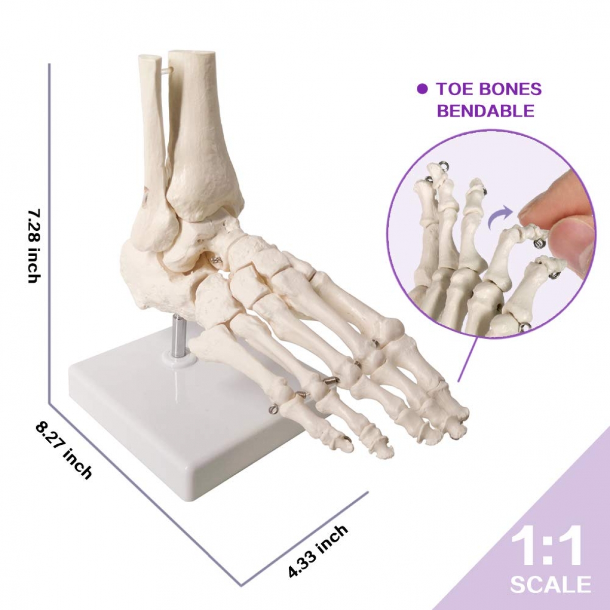
The metal hardware joints at the forefoot provide enhanced visual segmentation, showing how the toes articulate with the midfoot. This makes it especially useful for podiatric education, rehabilitation instruction, and orthotic consultations. Need to explain forefoot flexibility or the impact of a hallux valgus deformity? This model makes it straightforward.
Physiotherapists often ask: “Can I use this to show kinetic chain misalignments?” Yes. While the model is fixed, it’s ideal for explaining static alignment and how changes in foot structure can affect the knees, hips, and lumbar spine. It helps you tie localized anatomy into full-body biomechanics.
It’s highly popular across disciplines—used by orthopedic surgeons, sports injury therapists, anatomical lecturers, and kinesiologists. Whether you’re building a lesson plan on lower extremity motion or reviewing post-surgical foot structure with a patient, this model becomes the visual anchor you need.
Constructed from durable, lightweight synthetic resin, it’s designed for heavy use. The base-mounted display allows for stability during demonstrations and ease of transport. It’s equally at home on a teaching podium, consultation desk, or physiotherapy bench.
Semantic match is strong for search terms like “skeletal foot model,” “orthopedic foot joint tool,” “foot bone structure teaching model,” and “anatomical display for podiatry.” It stands out not just by design—but by its day-to-day performance in real-world education.
“This model simplified our approach to teaching foot biomechanics. It's become a staple in our podiatry courses,” shares Dr. Malik K., Academic Dean, Dubai Health Sciences Institute.
From diagnosis to demonstration, this foot model doesn’t just help you teach—it helps people understand, clearly and confidently.
The foot is often overlooked until something goes wrong—pain, misalignment, or reduced mobility. That’s why clear, structured education is essential, and the Life-Size Foot Joint Model helps deliver it. Whether you're walking a first-year med student through tarsal anatomy or helping a patient understand their recovery plan, this model supports your message with unmistakable clarity.
Its value lies not in complexity, but in its faithful representation of the core skeletal architecture of the foot and ankle. For learners, it brings textbook pages to life. For practitioners, it sharpens explanations. And for patients, it replaces confusion with comprehension. The result is improved engagement, retention, and trust—three pillars of effective health communication.
Frequently asked: “Is it only for podiatry?” Far from it. This model serves sports therapists, orthopedic clinics, rehabilitation centers, osteopaths, physiotherapists, and even footwear designers. Anywhere precise understanding of the foot’s skeletal framework is needed, this model provides the anatomical truth you can point to.
The neutral ivory tone and clean finish make it suitable for professional settings, while the size and sturdiness make it ideal for daily use. Mounted on a weighted base, it stays upright even during active discussion or multi-angle demonstration. No wobbles, no distractions—just reliable performance.
It continues to rank highly for search queries like “life-size foot skeleton,” “joint model for podiatric education,” “orthopedic foot bones model,” and “anatomical teaching aid for lower limb.” That’s because it delivers consistently on expectations—helping people see, touch, and understand.
“It’s a compact model, but it covers everything we need—students grasp alignment, clinicians explain injuries, and our patients get it faster,” notes Sneha D., Head Physiotherapist, Delhi.
Some tools serve a purpose. Others become part of your daily practice. The MYASKRO Foot Joint Model belongs in the latter category. It doesn’t ask for attention, but it earns respect. And in every setting where anatomy meets application, it quietly does its job—making complexity clear, one joint at a time.
Total Reviews (0)