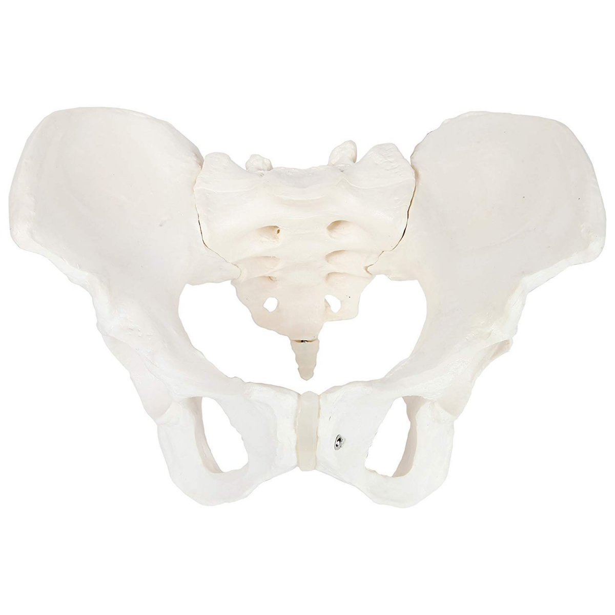
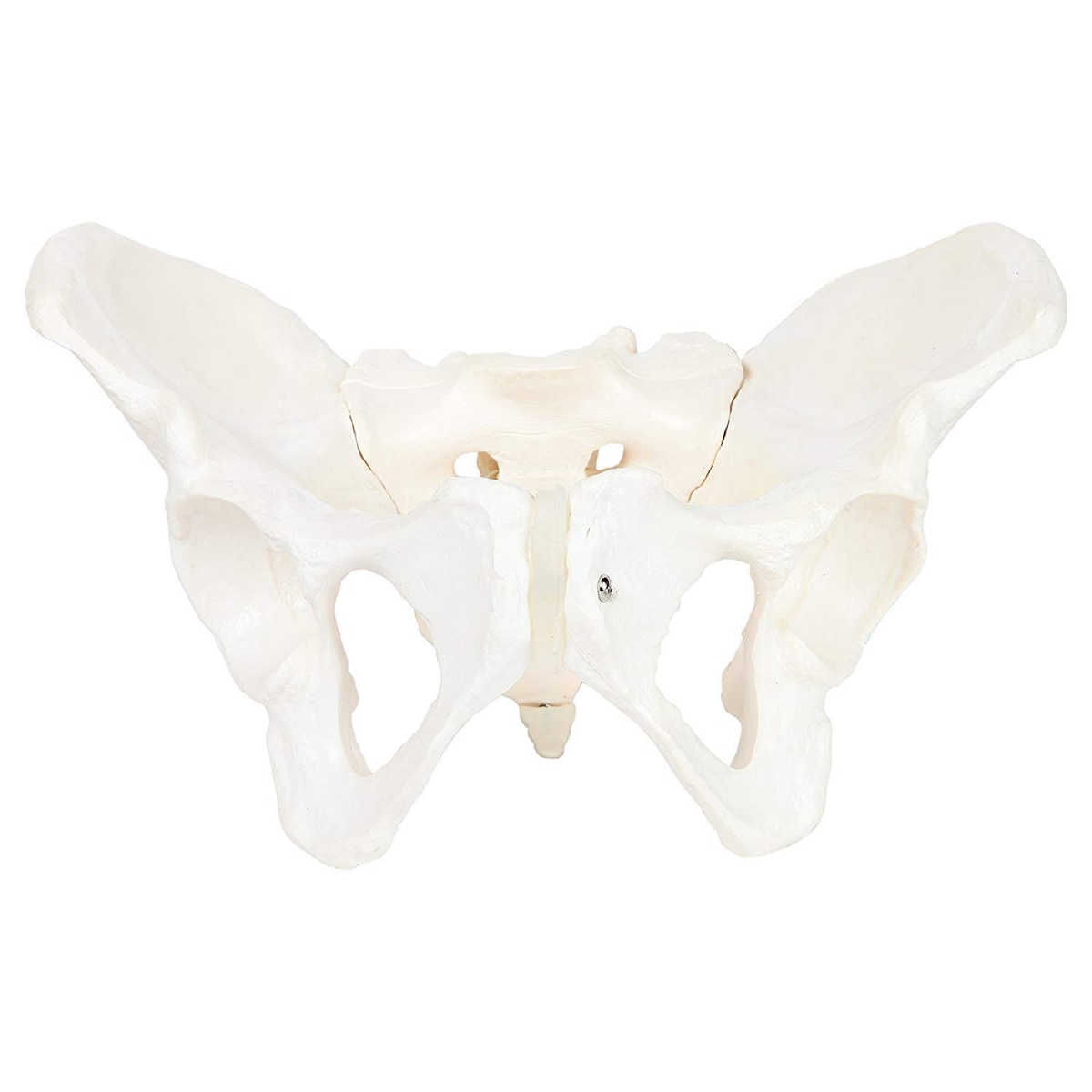
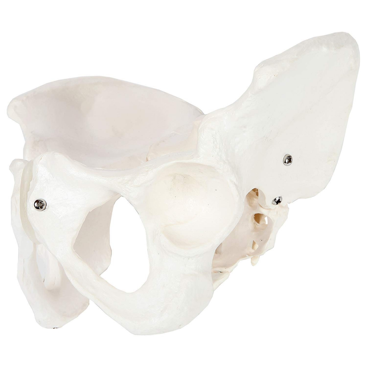
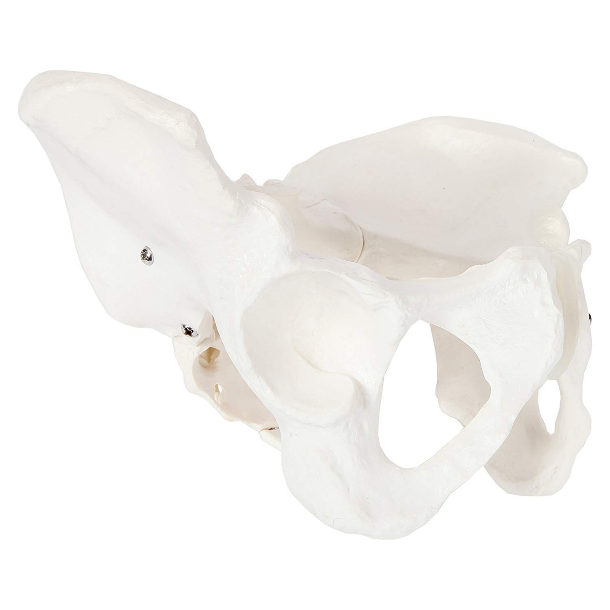
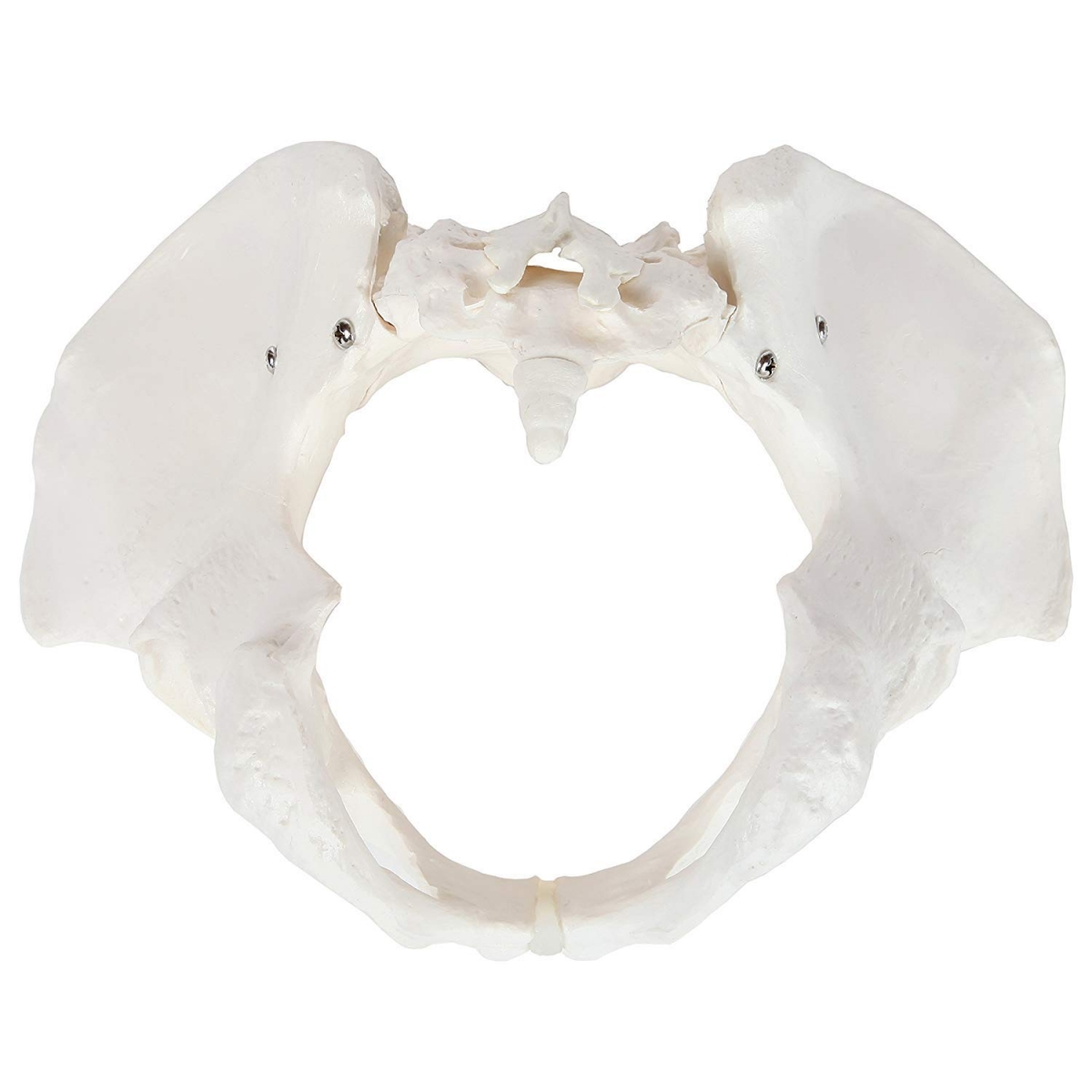
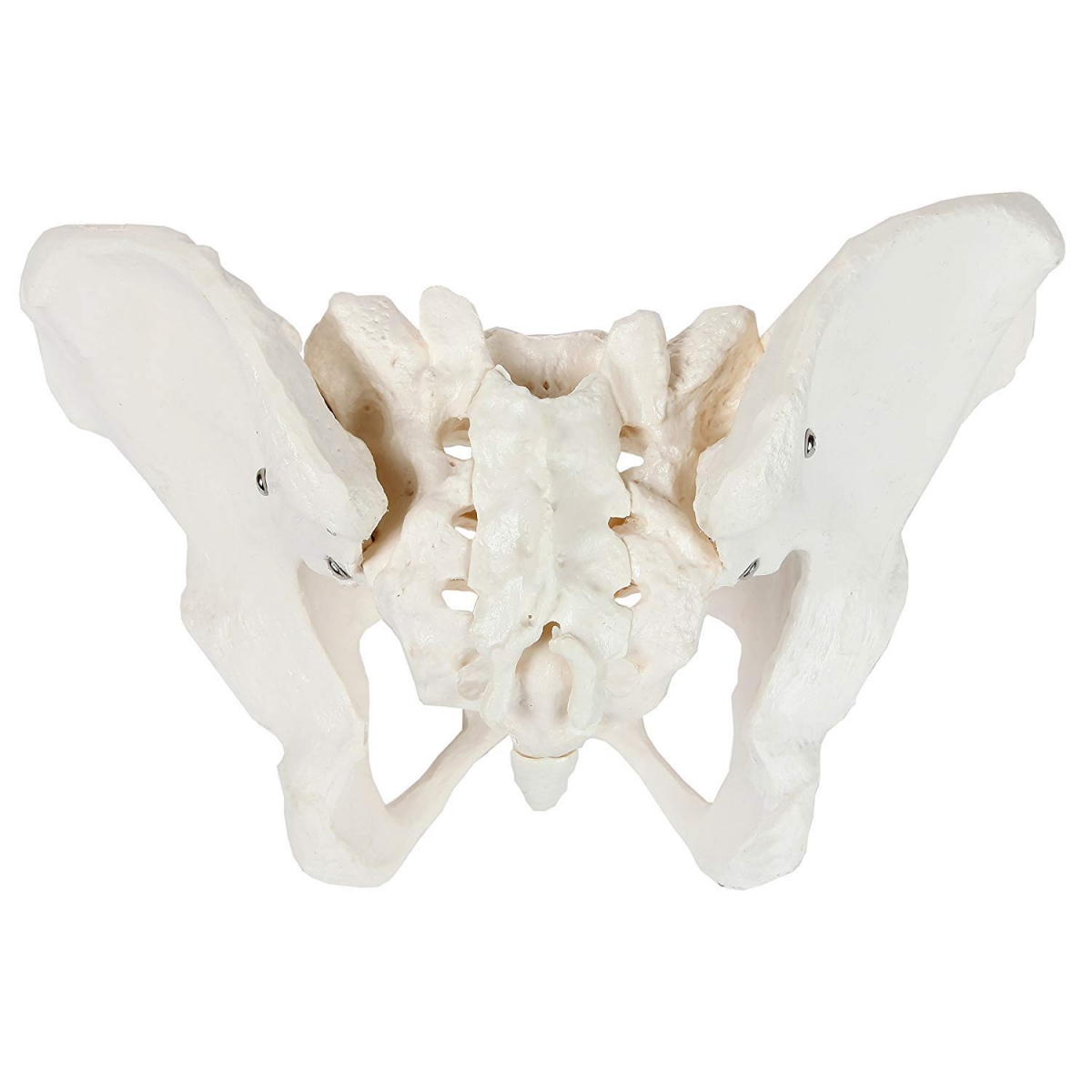
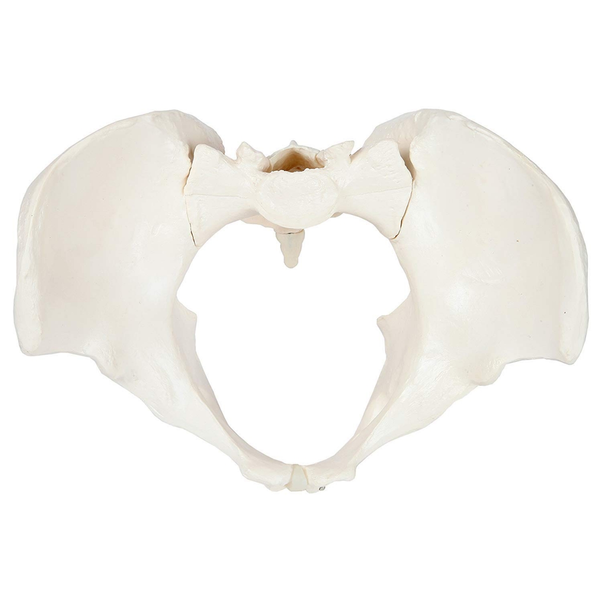
The MYASKRO Female Pelvis Anatomical Model with Sacrum and
Coccyx is a three-dimensional teaching model designed for studying
the bones, openings, joints and general proportions of the female bony
pelvis.
It displays the right and left hip bones together with the sacrum, coccyx
and pubic symphysis, allowing learners to understand how these structures
combine to form the complete pelvic ring.
The model is suitable for MBBS, nursing, physiotherapy, midwifery and
allied-health education, anatomy laboratory demonstrations, practical
examination preparation and introductory obstetric anatomy teaching.
Complete Female Bony Pelvis Model
The model presents the main components of the female pelvic skeleton in
their assembled anatomical relationship.
It shows:
- Right and left hip bones
- Ilium, ischium and pubis regions
- Sacrum
- Coccyx
- Pubic symphysis
- Right and left sacroiliac regions
- Right and left acetabula
- Obturator foramina
- Anterior and posterior sacral foramina
This complete arrangement gives learners a more useful three-dimensional
reference than studying individual pelvic bones separately.
Study the Ilium, Ischium and Pubis
Each hip bone is formed by three principal regions: the ilium, ischium and
pubis. The model helps students understand how these regions join around
the acetabulum and contribute to the walls of the pelvic cavity.
Visible landmarks and surfaces can be examined from different directions
to improve recognition during anatomy practicals and viva examinations.
Pelvic Inlet, Cavity and Outlet
When viewed from above, the model provides a clear view of the pelvic inlet
and the bony boundaries surrounding the pelvic cavity.
The inferior view helps learners study the pelvic outlet, pubic arch,
ischial regions and the relationship between the anterior and posterior
components of the pelvis.
The model can support the study of:
- Pelvic brim and inlet
- Greater and lesser pelvic regions
- Pelvic cavity
- Pelvic outlet
- Pubic arch and subpubic region
- Relationship between the sacrum and paired hip bones
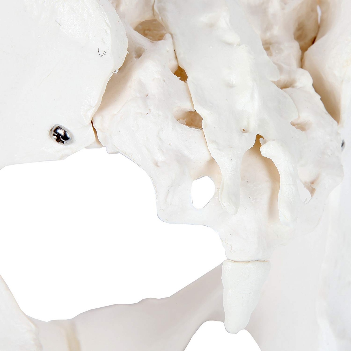
Sacrum, Coccyx and Sacroiliac Regions
The posterior portion of the model includes the sacrum and coccyx positioned
between the right and left hip bones.
The sacroiliac regions demonstrate where the ilium meets the sacrum, while
the anterior and posterior sacral foramina provide additional landmarks for
studying sacral anatomy.
Acetabula and Obturator Foramina
The right and left acetabula are shown on the lateral sides of the pelvis.
These cup-shaped sockets form the bony receiving surfaces of the hip joints.
The large obturator foramina can also be viewed clearly, helping students
understand their position between the pubic and ischial regions of each hip
bone.
Female Pelvic Anatomy
The model is designed to represent the general proportions of a female bony
pelvis. It can be used when introducing the wider pelvic opening, broader
pubic arch and pelvic cavity associated with female pelvic anatomy.
It is useful for comparing the general structural differences between male
and female pelvis models when both versions are available for classroom
demonstration.
This model is intended for anatomical education. It should not be used for
clinical measurement, obstetric assessment or diagnosis.
Multi-Angle Study and Demonstration
The model can be examined from anterior, posterior, superior, inferior and
lateral perspectives. This helps learners develop a three-dimensional
understanding of structures that may be difficult to interpret from a flat
textbook illustration.
Different views help demonstrate:
- Anterior pubic symphysis and pubic arch
- Posterior sacrum and sacral foramina
- Superior pelvic inlet
- Inferior pelvic outlet
- Lateral acetabular and iliac regions
- Internal pelvic surfaces
What Can Be Studied with This Model?
- Female bony pelvis anatomy
- Pelvic girdle organisation
- Ilium, ischium and pubis identification
- Sacrum and coccyx orientation
- Pubic symphysis
- Sacroiliac regions
- Pelvic inlet and outlet
- Acetabula and hip-joint orientation
- Obturator foramina
- Basic obstetric pelvic anatomy
Who Should Buy This Female Pelvis Model?
The model is suitable for:
- MBBS and medical students
- Nursing students and colleges
- Physiotherapy students
- Midwifery and obstetric training programmes
- Occupational therapy students
- Allied-health science students
- Anatomy teachers and tutors
- Medical and nursing skill laboratories
- Students preparing for practical and viva examinations
Why Choose the MYASKRO Female Pelvis Model?
Complete pelvic ring: Shows both hip bones, sacrum, coccyx
and pubic symphysis in their assembled relationship.
Female anatomical proportions: Designed for teaching the
general structure and proportions of the female bony pelvis.
Multiple visible landmarks: Displays the acetabula,
obturator foramina, sacral foramina and sacroiliac regions.
Multi-angle learning: Can be examined from superior,
inferior, anterior, posterior and lateral perspectives.
Useful for practical revision: Supports three-dimensional
orientation, bone identification and viva preparation.
Institutional purchase support: GST invoice, formal
quotation, proforma invoice and bulk-order assistance are available for
colleges and hospitals.
Product Specifications
|
Product Name
|
MYASKRO Female Pelvis Anatomical Model
|
|
Brand
|
MYASKRO
|
|
Product Type
|
Female bony pelvis anatomical model
|
|
Anatomical Representation
|
Female pelvis
|
|
Main Components
|
Two hip bones, sacrum, coccyx and pubic symphysis
|
|
Hip Bone Regions
|
Ilium, ischium and pubis
|
|
Displayed Landmarks
|
Acetabula, obturator foramina, sacral foramina, pelvic inlet and outlet
|
|
Colour
|
Natural bone colour
|
|
Primary Use
|
Anatomy, pelvic girdle and introductory obstetric teaching
|
|
Recommended For
|
Medical, nursing, physiotherapy and midwifery education
|
Note: Colour, surface finish, dimensions and minor
construction details may vary slightly between production batches without
affecting the educational purpose of the model.
Frequently Asked Questions
Is this a female pelvis model?
Yes. It is designed to represent the general bony proportions and structure
of a female human pelvis.
What bones are included?
The model includes the right and left hip bones, sacrum and coccyx, joined
anteriorly at the pubic symphysis.
Does it show the pelvic inlet and outlet?
Yes. The pelvic inlet can be viewed from above, while the pelvic outlet and
pubic arch can be examined from the inferior side.
Are the acetabula visible?
Yes. Both hip sockets or acetabula are displayed on the lateral sides of the
pelvis.
Does the model include the sacrum and coccyx?
Yes. The sacrum and coccyx are included in the posterior portion of the
pelvic model.
Is it suitable for obstetric teaching?
It is suitable for introducing female bony-pelvis anatomy, pelvic inlet and
outlet orientation and general obstetric anatomical concepts. It is not
intended for clinical pelvimetry or diagnosis.
Is the model detachable?
The photographed model is assembled using visible fittings. Do not advertise
it as detachable unless separation and reassembly have been physically
confirmed for the supplied product.
Is it suitable for physiotherapy students?
Yes. It can support the study of pelvic anatomy, sacroiliac regions,
acetabula and general pelvic orientation.
Can institutions request a formal quotation?
Yes. Colleges, hospitals and training institutions can request a formal
quotation, proforma invoice, GST documentation and estimated dispatch
schedule.