

Components
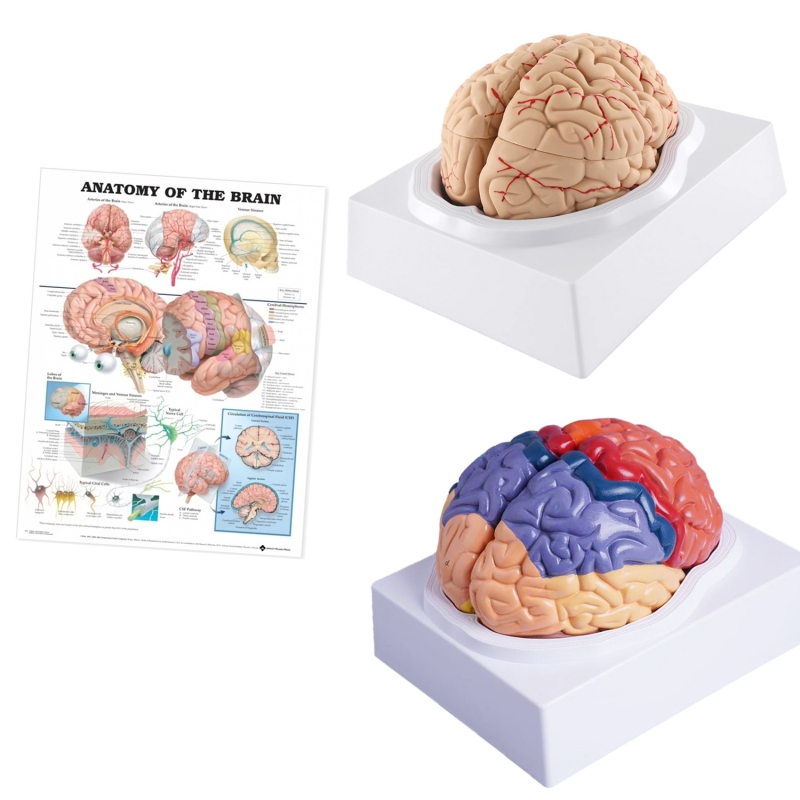
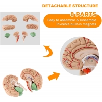
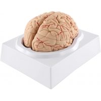
Human Brain Model (8 removable parts): Right/left cerebral hemispheres with detachable frontal, parietal, temporal, occipital lobes; cerebellum; brainstem (midbrain–pons–medulla). Keyed assembly on base.
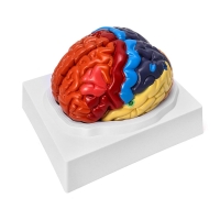
Brain Model with Painted Cortical Regions: Color-differentiated lobes with major functional areas approximated—precentral (primary motor), postcentral (primary somatosensory), Broca, Wernicke, primary visual (V1), primary auditory (A1)—for rapid orientation.
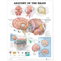
Brain Anatomy Chart (52 × 70 cm, special lamination with rollers): Lateral/medial/inferior views, cranial fossae, ventricular system, and vascular overview.
Learning objectives
Identify lobes, central sulcus, lateral (Sylvian) fissure, parieto-occipital sulcus, cerebellar folia, and brainstem segments.
Map cortical regions to function and common deficits (e.g., contralateral hemiparesis—precentral; receptive aphasia—posterior superior temporal; homonymous hemianopia—occipital cortex).
Orient to limbic structures (cingulate gyrus/hippocampal formation—schematic) and ventricular system for CSF pathway discussion.
Use in OSCEs for lobe identification, assembly/disassembly under time, and stroke-territory correlation (ACA/MCA/PCA at a teaching level).
Specifications
Scale: life-size; material: Medical-Grade PVC.
Mounting: individual stable bases; parts are magnetic/press-fit.
Chart: heavy-gauge lamination, dry-wipe compatible with top–bottom rollers.
Care: wipe with mild detergent or 70% alcohol; avoid solvents/heat.
Intended use: instructional aids for MBBS/BDS, physiotherapy, psychiatry/neurology teaching, and skill labs.
Total Reviews (0)