


 | MYASKRO")
₹ 3,199 ₹ 4,895
")
₹ 2,899 ₹ 10,579

₹ 7,999 ₹ 22,379
 - Myaskro")
₹ 21,899 ₹ 34,179
")
₹ 9,599 ₹ 22,379

₹ 1,899 ₹ 3,417

₹ 3,899 ₹ 11,641

₹ 1,699 ₹ 3,371

₹ 5,599 ₹ 9,622

₹ 2,199 ₹ 5,191
 With Special Lamination")
₹ 1,599 ₹ 3,865

₹ 4,699 ₹ 10,083
 - MYASKRO")
₹ 2,499 ₹ 9,021
 3-Part – Ideal for MBBS, BDS & Medical Education")
₹ 2,899 ₹ 6,189
 - MYASKRO")
₹ 4,699 ₹ 8,431

₹ 2,949 ₹ 10,579
 Life Size")
₹ 1,699 ₹ 4,865

₹ 5,499 ₹ 9,268
")
₹ 14,899 ₹ 21,116

₹ 14,899 ₹ 25,836

₹ 19,599 ₹ 25,801
 | Myaskro")
₹ 3,199 ₹ 4,865
From sutures to sulci—teach neuroanatomy with clinical clarity.
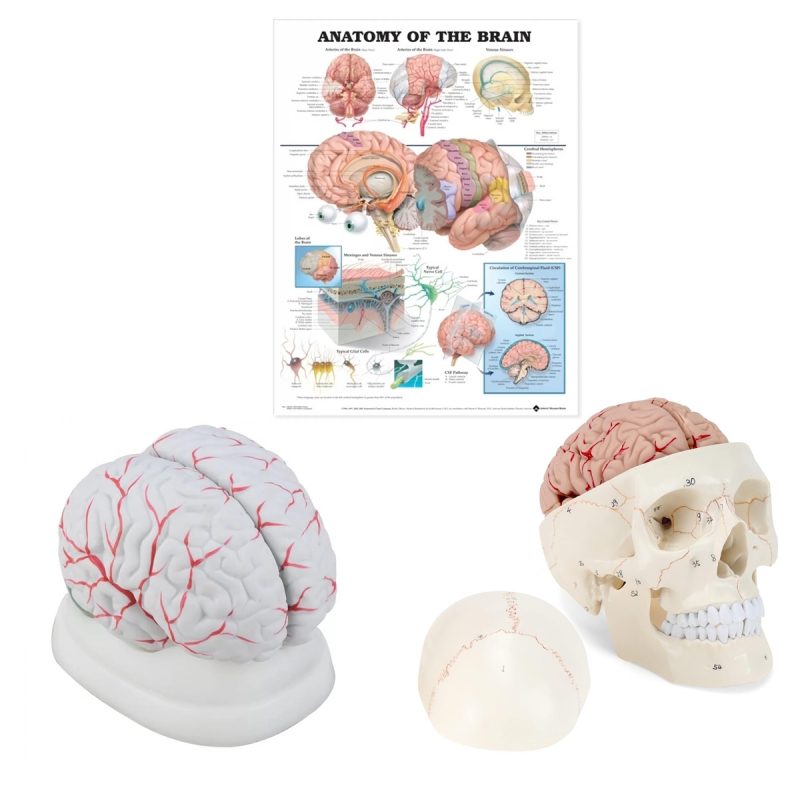
A three-piece set that aligns skull landmarks with cortical anatomy and core neuro concepts. Perfect for MBBS/MD, BSc Nursing, PT/OT programs, neurology & neurosurgery OPDs, and OSCE/viva prep.
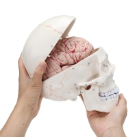
Numbered Skull with 8-Part Brain (fits inside the cranium)
Removable calvaria; clearly numbered bones, sutures, and foramina for cranial base orientation.
8-piece brain segmentation exposes cortical lobes, cerebellum, and brainstem for tract, lesion-localisation, and hemisphere teaching.
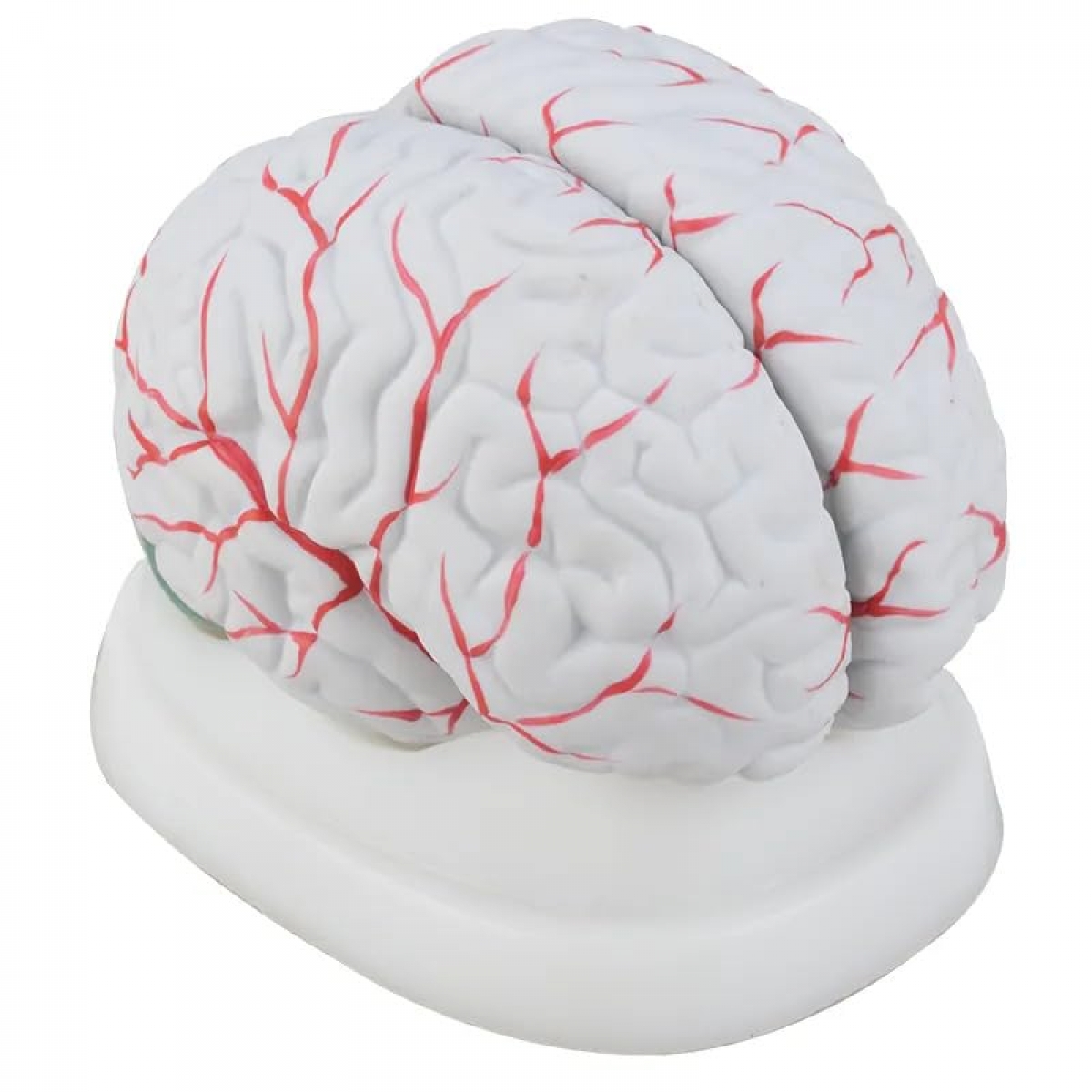
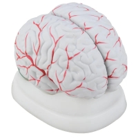
8-Part Brain on Stand
Stand-mounted for bench demonstrations; external gyri/sulci and superficial cerebral vasculature highlighted to discuss arterial territories and stroke patterns.
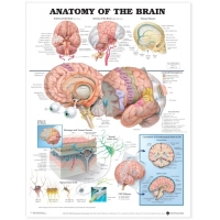
Brain Anatomy Chart – 52 × 70 cm (special lamination with rollers)
Clinic-ready wall chart covering lobes & functional areas, meninges/dural folds, ventricles & CSF circulation, major arteries/venous sinuses, and key cross-sections.
Wipe-clean lamination with rollers for easy hanging and transport.
Link structure to function: pair numbered foramina with cranial nerve pathways; relate cortical areas to deficits.
Explain pathology fast: stroke territories, mass effect, raised ICP, meningitis/CSF flow blockage—shown with models and chart together.
Built for daily use: durable PVC/resin, stable bases, and easy-clean surfaces for OPD, skills labs, and community sessions.
Total Reviews (0)