

Anatomic Details
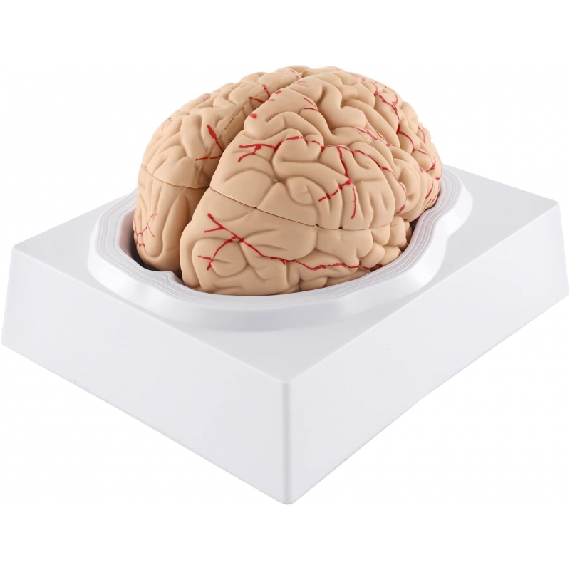
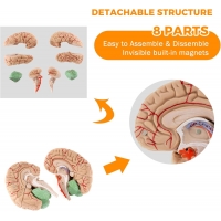
Right and left cerebral hemispheres with detachable lobar components (frontal, parietal, temporal, occipital); clear gyri/sulci relief and superficial cortical vessels.
Cerebellum (hemispheres with vermis) and brainstem (midbrain–pons–medulla) as separate pieces.
Median section shows corpus callosum, thalamus, hypothalamus, ventricular cavity outline, and basilar/circle-of-Willis vessels (schematic).
Key learning outcomes
Lobar identification and surface landmarking (central sulcus, lateral fissure, parieto-occipital sulcus).
Correlation of cortical regions with function (motor/somatosensory, language, visual, auditory).
Orientation to cerebellar anatomy and brainstem levels for cranial-nerve localisation.
Construction
8-piece model with hidden magnets/keyed joints for secure assembly.
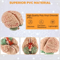
Medical-grade PVC, matte finish; resistant to routine disinfection.
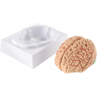
Stabilising cradle-base for bench or OSCE station use.
Use
UG/PG neuroanatomy, neurology/psychiatry teaching, physiotherapy; OSCE/viva (lobe ID, midline anatomy).
Patient education for stroke, tumour, and TBI topography.
Total Reviews (0)