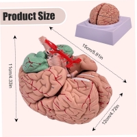


Anatomic Details
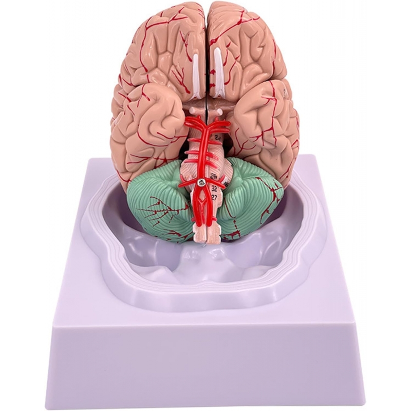
Bilateral cerebral hemispheres (frontal, parietal, temporal, occipital lobes demarcated); crisp gyri/sulci and superficial cortical vessels.
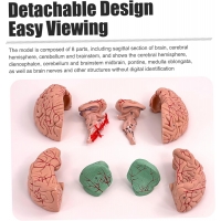
Cerebellum (two hemispheres with vermis) and brainstem split into upper (midbrain–pons) and lower (medulla) segments.
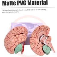
Median section shows corpus callosum, diencephalon (thalamus/hypothalamus), third/fourth ventricles, and schematic basilar/Circle of Willis arteries.
Educational use
Lobar identification; surface landmarking (central sulcus, lateral fissure, parieto-occipital sulcus).
Functional correlation (motor/somatosensory cortices, language areas, visual/auditory cortices).
Orientation to cerebellar topography and brainstem levels for cranial-nerve localization; OSCE assembly/disassembly.
Construction / specifications
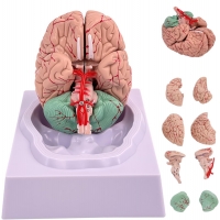
8 detachable parts with hidden magnetic/keyed joints; numbered landmarks.
Matte medical-grade PVC; wipe-clean, classroom-durable.
Cradle display base. Approx. size 15 × 12 × 11 cm.
Care
Clean with mild detergent or 70% alcohol; avoid solvents/heat.
Intended use
Instructional model for neuroanatomy, neurology, psychiatry, physiotherapy teaching and patient education.
Total Reviews (0)