

Composition
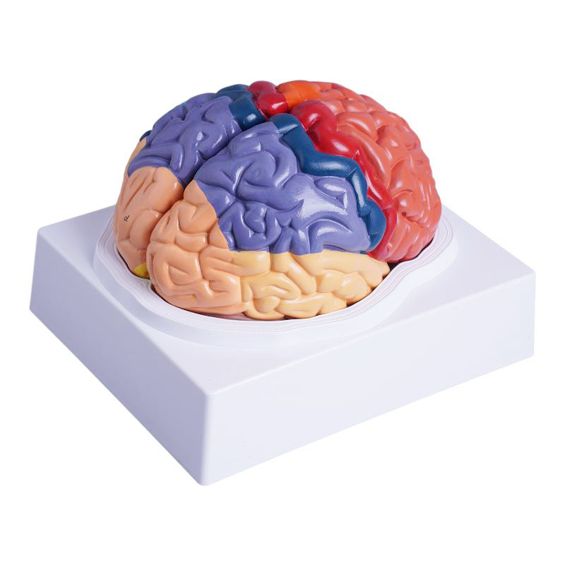
Bilateral cerebral hemispheres mounted in a cradle base; color segmentation of frontal, parietal, temporal, occipital lobes.
Superficial arterial markings and clear relief of gyri and sulci along the longitudinal fissure.
Morphologic detail
High-contrast demarcation of key areas for rapid teaching: precentral (primary motor) gyrus, postcentral (primary somatosensory) gyrus, Broca and Wernicke regions (approximate), primary visual (V1), primary auditory (A1).
Prominent central sulcus, lateral (Sylvian) fissure, and lobar boundaries.
Educational use
Lobar identification, cortical function mapping, and bedside correlation of deficits (e.g., hemiparesis—precentral; receptive aphasia—posterior superior temporal; hemianopia—occipital).
Suitable for UG/PG neuroanatomy, neurology/psychiatry tutorials, speech-language teaching, and OSCE spotters.
Specifications
Scale: life-size • Material: Medical-Grade PVC, matte finish • Mount: stable base (removable brain).
Cleaning: wipe with mild detergent or 70% alcohol; avoid solvents/heat.
Intended use: instructional model;
Key Highlights
LIFE-SIZE, COLOR-CODED LOBES for instant cortical orientation.
CRISP GYRI/SULCI with superficial arterial tracings for landmarking.
FUNCTIONAL AREAS HIGHLIGHTED (motor, sensory, language, visual, auditory).
DURABLE PVC — classroom-tough, wipe-clean.
OSCE-READY — ideal for rapid identification and viva explanations.
Total Reviews (0)