

Anatomical Details
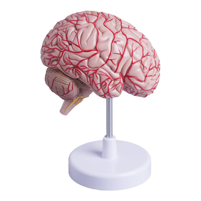
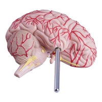
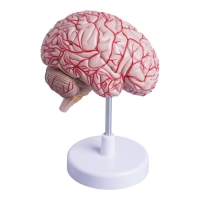
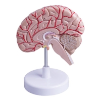
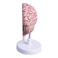
Single hemispheric cast on stand; external surface with pronounced gyri/sulci and painted superficial cerebral arteries.
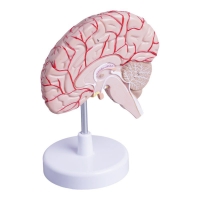
True median (sagittal) section showing corpus callosum, cingulate gyrus, septum pellucidum/ventricular outline (lateral & third), thalamus–hypothalamus, midbrain–pons–medulla, and cerebellum with arbor vitae.
Brainstem displays root-entry/exit zones of selected cranial nerves.
Educational use
Rapid lobar identification and landmarking: central sulcus, lateral (Sylvian) fissure, parieto-occipital and calcarine sulci.
Correlate ACA/MCA/PCA superficial territories with focal neurological deficits.
Orient to commissural, diencephalic, and brainstem–cerebellar relationships for cranial-nerve localisation and CSF pathway discussion.
Suitable for UG/PG neuroanatomy, neurology/psychiatry tutorials, SLP teaching, and OSCE spotters.
Specifications
Scale: life-size half brain (single piece).
Material: matte, medical-grade PVC; hand-painted vessels.
Mount: removable model on round base/rod.
Cleaning: wipe with mild detergent or 70% alcohol; avoid solvents/heat.
Total Reviews (0)