

Anatomical Details
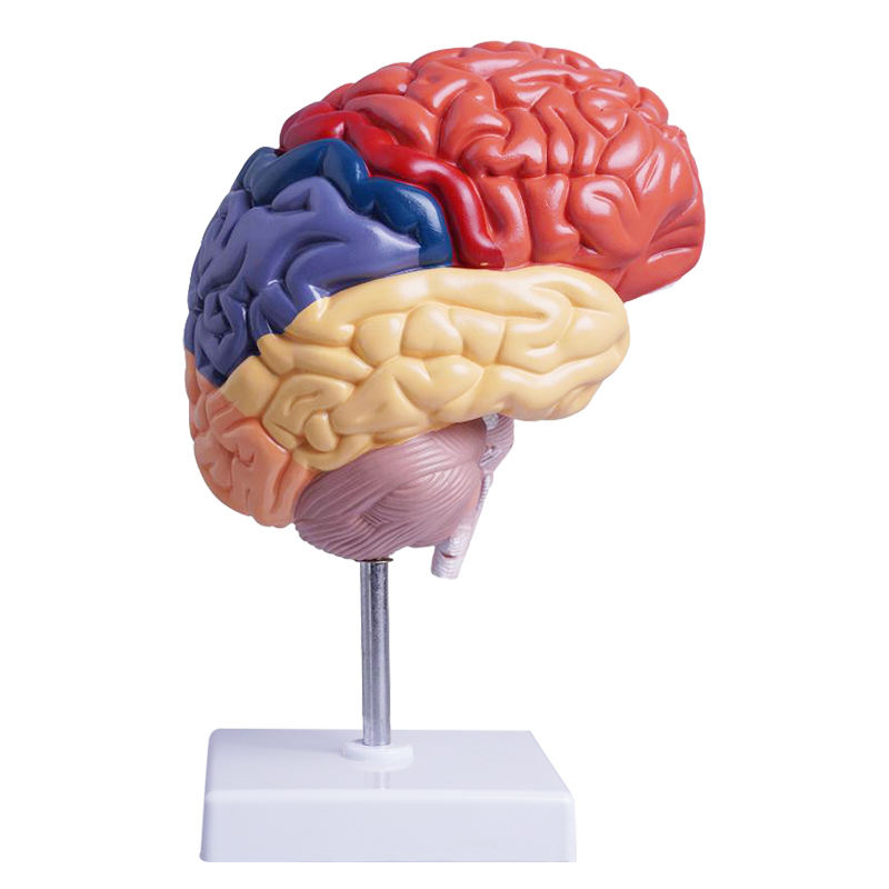
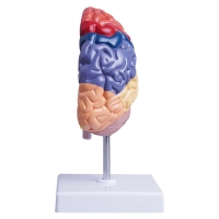
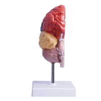
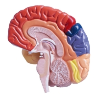
Single hemispheric cast mounted on stand; color-segmented lobes: frontal, parietal, temporal, occipital.
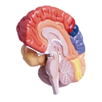
True median (sagittal) section displaying corpus callosum, cingulate gyrus, diencephalon (thalamus/hypothalamus), midbrain–pons–medulla, and cerebellum with arbor vitae.
Clear relief of gyri and sulci; superficial arterial tracings.
Educational use
Rapid lobe identification and correlation with function (motor/somatosensory, language, visual, auditory).
Orientation to limbic structures, commissural fibers, and ventricular outlines (lateral/third ventricles).
Brainstem–cerebellar relationships for cranial-nerve localization discussions.
Ideal for UG/PG neuroanatomy, neurology/psychiatry tutorials, SLP teaching, and OSCE spotters.
Specifications
Scale: life-size half brain.
Material: medical-grade PVC, matte finish, hand-painted; labeled regions.
Mount: removable model on stable base/rod.
Care: wipe with mild detergent or 70% alcohol; avoid solvents/heat.
Key Highlights
LIFE-SIZE COLORED LOBES for instant cortical orientation.
MEDIAL SECTION DETAIL: corpus callosum, diencephalon, ventricles, brainstem, cerebellum.
CRISP GYRI/SULCI with vascular tracings for dependable landmarking.
DURABLE PVC—classroom-tough, wipe-clean.
OSCE-READY—compact stand for station use.
Total Reviews (0)