
₹ 2,199 ₹ 5,191

₹ 4,699 ₹ 10,083

₹ 7,999 ₹ 22,379
")
₹ 9,599 ₹ 22,379
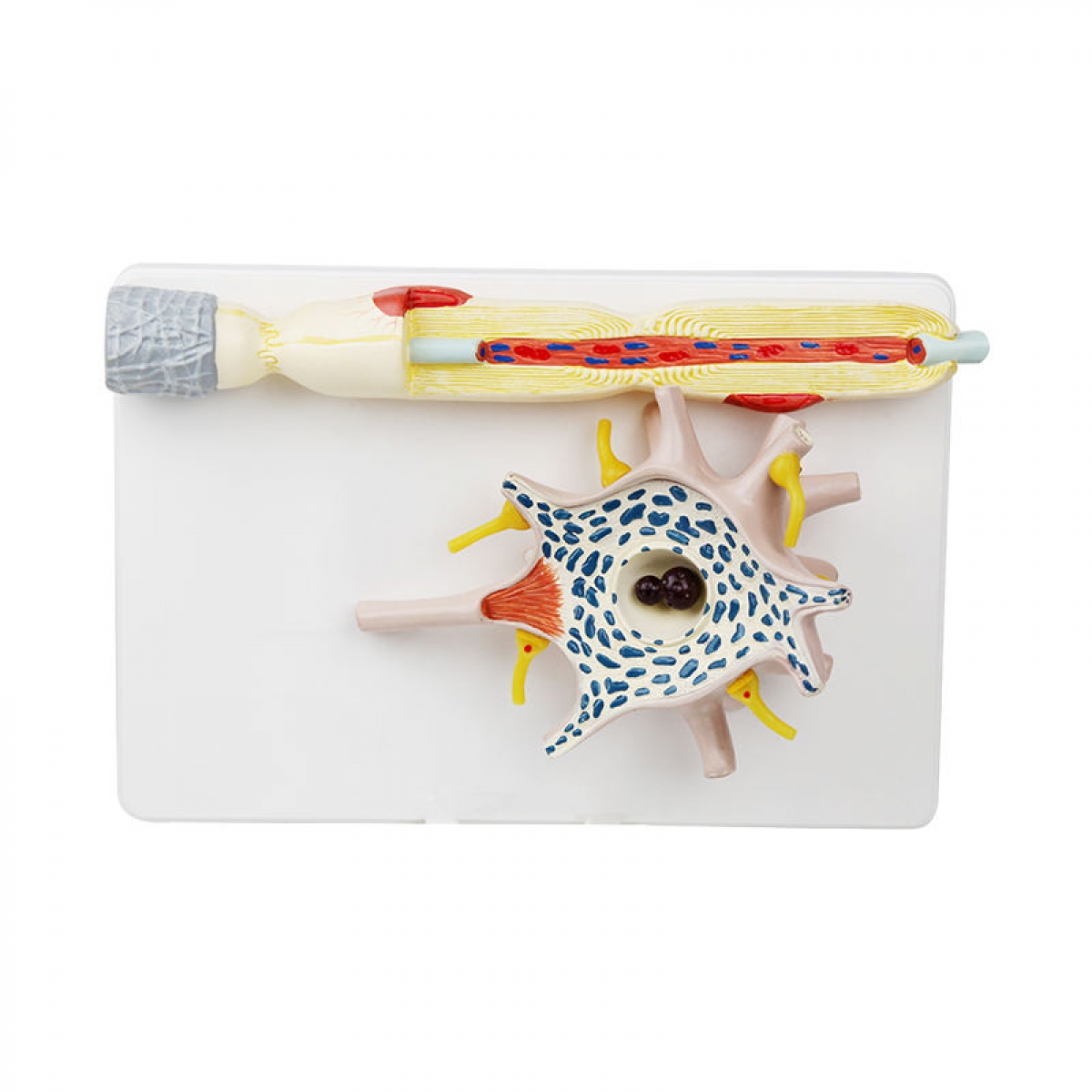
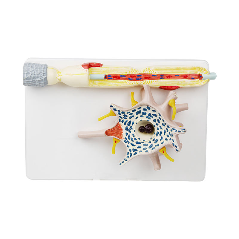
At Anatomy Shop, we understand that mastering neuroanatomy requires more than two-dimensional illustrations. Our Neuron Model is engineered to provide a comprehensive, three-dimensional perspective on the microanatomy and functional architecture of the nerve cell.
This highly enlarged model captures the intricate details of a multipolar neuron, including:
Cell Body (Soma): Clearly demonstrates the nucleus, prominent nucleolus, Nissl granules, and perikaryon—critical for discussions on neuronal metabolism and protein synthesis.
Dendritic Arborization: Extensive branching to visualize afferent signal reception, facilitating explanation of synaptic input and integration.
Axon Hillock and Myelinated Axon: Distinct axon hillock for teaching action potential initiation; myelin sheath, nodes of Ranvier, Schwann cells, and internodal segments depicted for illustrating saltatory conduction.
Axon Terminals & Synaptic Endings: Highlighting synaptic boutons and pre-synaptic structures—ideal for explaining neurotransmission, synaptic plasticity, and neuropharmacology.
Vascular and Supporting Elements: Includes representations of neuroglia and associated blood vessels for a holistic understanding of the neuronal microenvironment.
Clinical & Academic Applications:
Essential for explaining the structural basis of neurological pathologies such as demyelinating diseases (e.g., Multiple Sclerosis), neuropathies, and neurodegenerative conditions.
Supports neurophysiology, histology, and clinical neurology teaching at undergraduate and postgraduate levels.
Model Features:
Enlarged for high-visibility demonstration in lecture halls, practical labs, and clinics.
Durable, medical-grade construction—engineered for years of repeated use and tactile exploration.
Ideal for medical, dental, nursing, and allied health education.
At Anatomy Shop, our commitment is to anatomical excellence—delivering models that translate microscopic complexity into real-world clarity for future clinicians and educators.
Total Reviews (0)