Offer


Click to zoom
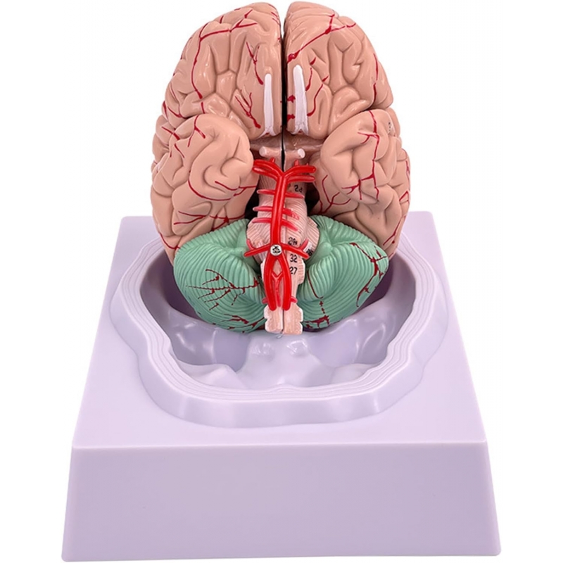


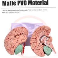
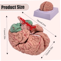
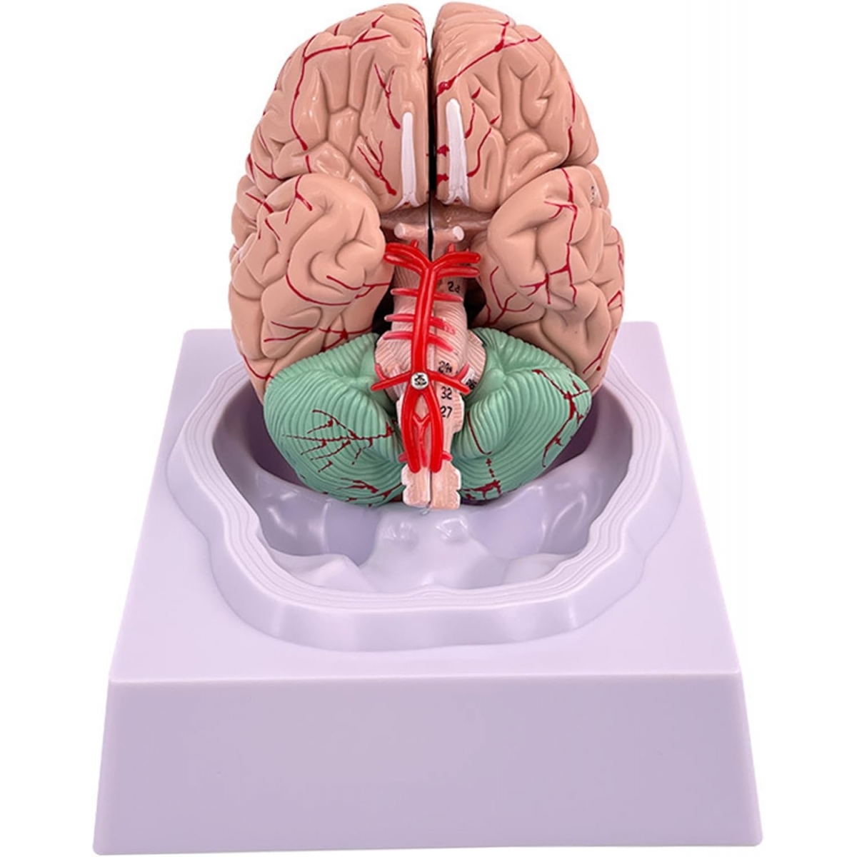
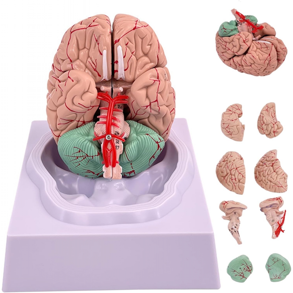
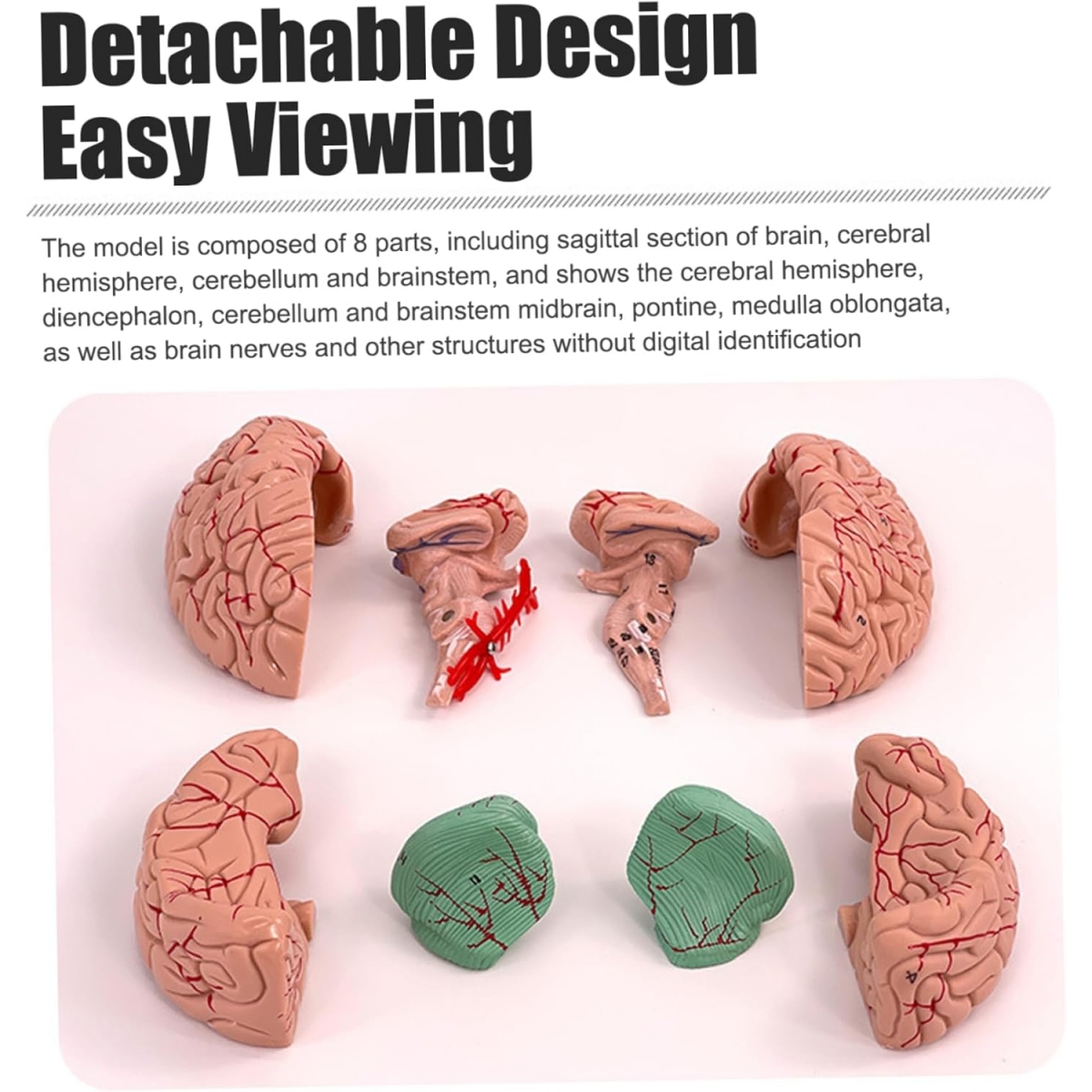
Premium Human Brain Model (Removable into 8-Parts) - Myaskro
₹ 21,899
₹ 34,179
You Save : ₹ 10,407
2 - 4 Working Days Delivery
In Stock
Secure Payment
3-Day Replacement
GST Invoice Available
Need GST quotation, bulk pricing or purchase support?
Get quotation on letterhead with product list, quantities and delivery details.
Get Quote / Bulk Pricing


")
")