57% OFF
Click to zoom
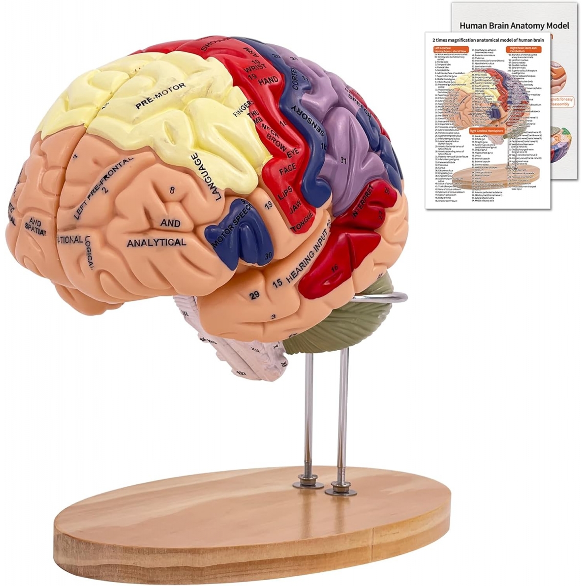
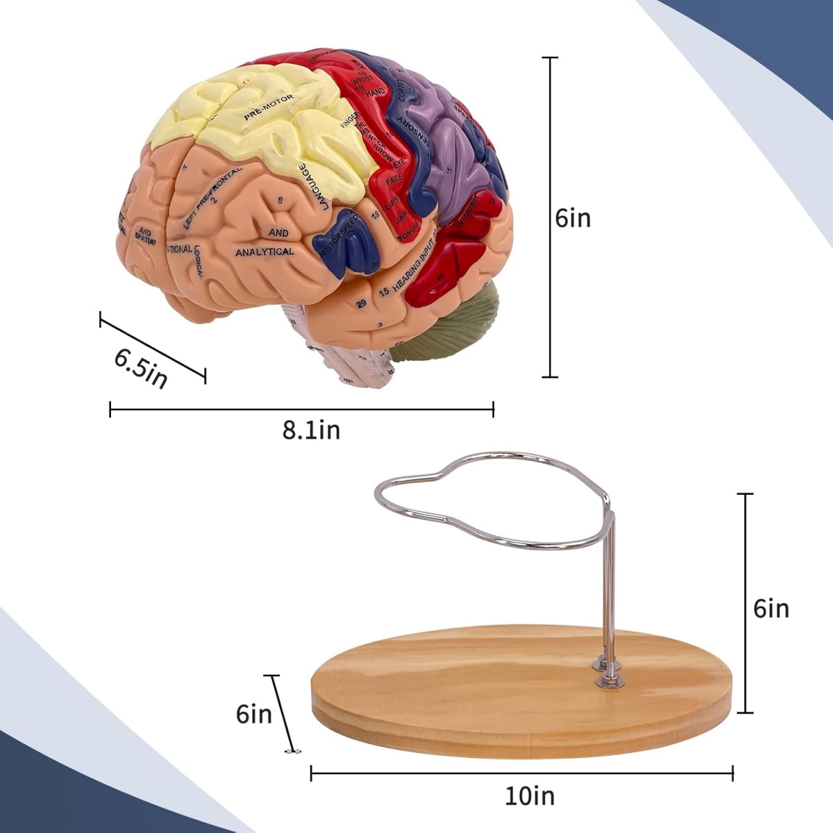
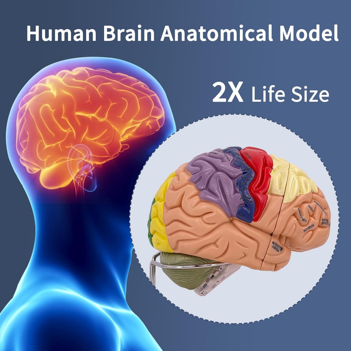
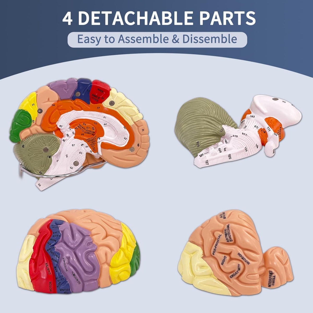
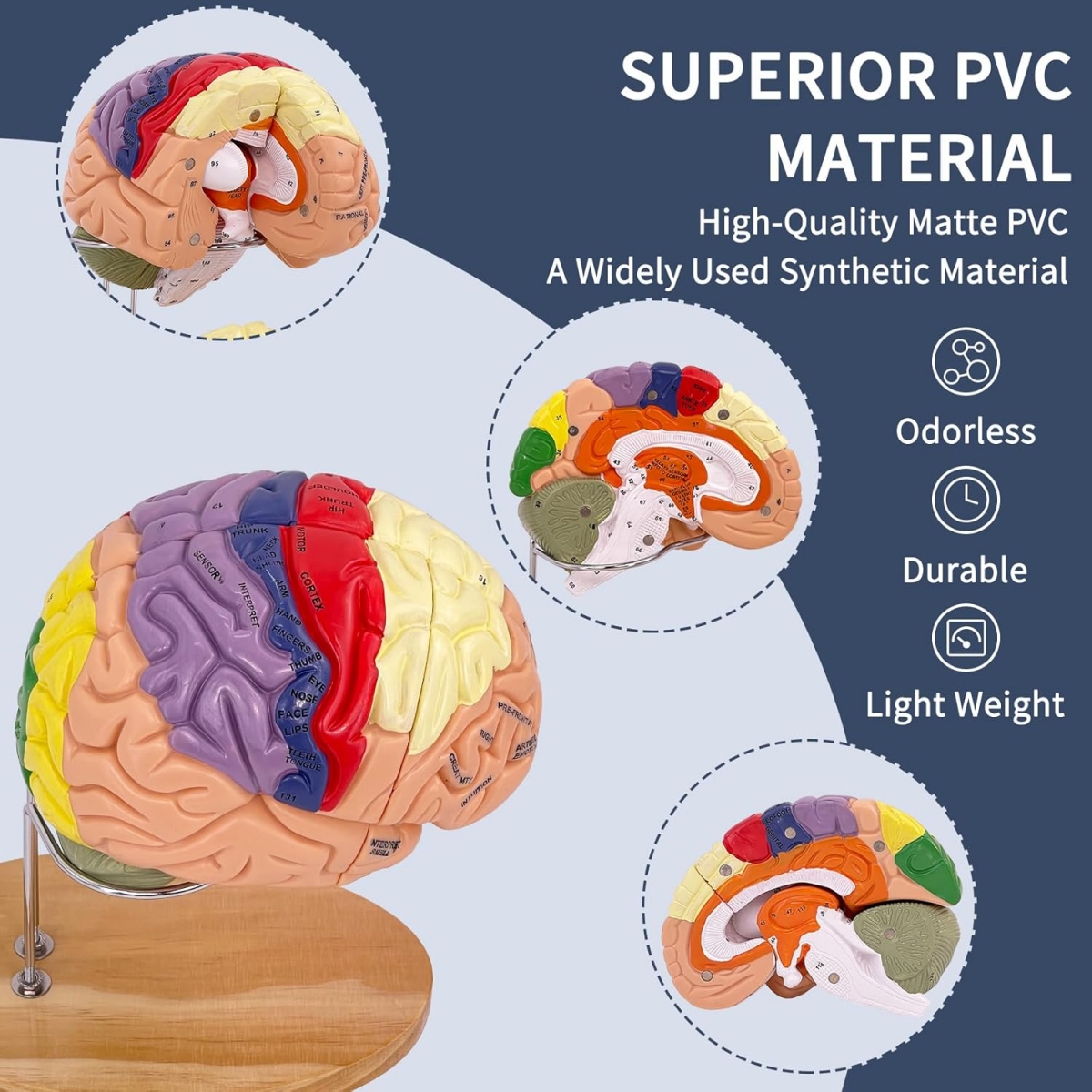
2 Times Enlarged Human Brain Model — MYASKRO (4 detachable parts)
INSTITUTIONAL LIST PRICE
₹ 9,599
₹ 22,379
57% OFF
Inclusive of GST & shipping
Final quote may vary based on quantity, delivery location and purchase requirement.
Pricing on Request
Get a tailored quote based on your requirements.
Timeline confirmed before dispatch
Institutional documentation support
GST invoice / proforma available
Request a Quote
Share your requirements
Get a personalized quote
Fast response from our team
2 - 4 Working Days Delivery
In Stock
Institutional Purchase Support
For colleges, hospitals, labs and training centres requiring official quotation, GST documentation and dispatch coordination.
We’ll respond by email or phone. WhatsApp support is optional after submitting.
Your information is secure and confidential.
Secure Payment
3-Day Replacement
GST Invoice Available
Need purchase documentation?
Share your requirement for an official quotation, proforma invoice and delivery timeline confirmation.
Request Purchase Support
 - Myaskro")


