MYASKRO® Transparent Lung, Trachea & Bronchial Tree With Heart – Premium Anatomical Teaching Model
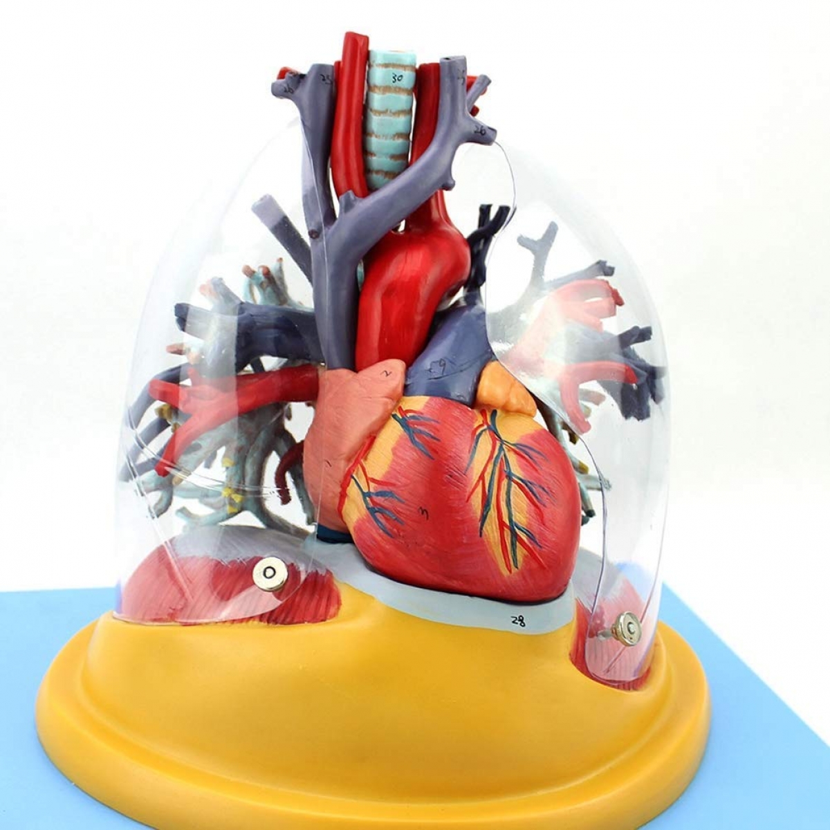
See Respiratory and Cardiac Anatomy in a Whole New Light – Literally.
The MYASKRO® Transparent Lung and Heart Model is a state-of-the-art educational tool combining the clarity of transparent materials with the precision of anatomical detail. This model is designed to give students, instructors, and medical professionals a full 3D view of the interconnected structure of the lungs, trachea, bronchial tree, and heart—all enclosed within a durable, transparent dome for 360° visibility.
It’s perfect for demonstrating the integrated function of the respiratory and circulatory systems, making it ideal for classroom instruction, clinical education, OSCE preparation, and patient consultation.
Key Features
- Transparent Lung Dome: Clearly see bronchial branches, pulmonary arteries, and airways
- Detailed Heart Model: Centrally located, the heart is color-coded and shows external vasculature
- Full Bronchial Tree: Includes trachea, left and right bronchi, and secondary branches
- Rigid Display Case: Protects components while allowing full model visibility from all angles
- Color-Coded Anatomy: Veins, arteries, and structural regions marked for instant recognition
What You Can Study
This combined anatomical model allows in-depth study of:
- Lungs: Upper, middle, and lower lobes of the left and right lungs
- Trachea & Bronchi: Branching structure of the airways including segmental bronchi
- Heart: External features, coronary vessels, and chamber outlines
- Vascular Network: Pulmonary arteries and veins entering and exiting the lungs and heart
The transparent design makes it easy to demonstrate how air moves through the respiratory system and how the heart and lungs interact during oxygen exchange.
Who It’s For
- Medical and Nursing Students: Gain a better understanding of the thoracic cavity’s organ systems
- Respiratory Therapists and Pulmonologists: Use as a teaching aid for airway function and disease
- Cardiologists: Show heart-lung interaction in circulatory education
- Biology Teachers: Demonstrate major systems in high school and college classrooms
- Patient Educators: Visually explain chronic lung conditions, surgeries, or cardiac interventions
Educational Advantages
- Integrated Design: Teaches two systems—respiratory and circulatory—in one interactive unit
- Better Engagement: Keeps students focused through transparent visualization
- 360-Degree Learning: No blind spots—view every side of the lungs and heart simultaneously
- Perfect for Demonstrations: Easily portable and durable enough for frequent use in labs and lectures
Materials and Construction
- Material: Medical-grade PVC with transparent polycarbonate dome
- Finish: Hand-painted anatomical structures with fade-resistant color
- Mount: Secure base with fixed internal support system
- Dimensions: Compact, desktop-friendly size ideal for classroom desks and doctor’s offices
Frequently Asked Questions
Is this a life-size model?
No. This model is slightly scaled down for compact display purposes but retains high anatomical accuracy and detail across all major thoracic structures.
What is the dome made of?
The transparent casing is crafted from durable, medical-grade polycarbonate, which is resistant to scratches, yellowing, and breakage—ideal for both transport and long-term classroom use.
Can the internal components be removed?
No. This is a non-dissectible, sealed display model designed for clear visibility rather than part separation. It provides a safe, static presentation for repeated handling.
Is this model labeled?
Yes. Major anatomical structures are number-coded and color-differentiated. A printed reference key (included or downloadable) is typically supplied for matching part numbers to names.
Clinical and Educational Use Cases
- Classroom Demonstrations: Engage students with an interactive, 3D overview of pulmonary and cardiac structure
- OSCE Prep: Teach and quiz students on the path and branching of airways or vascular entry/exit points
- Hospital Education Rooms: Visual aid for patient briefings on heart-lung surgeries or respiratory disorders
- Lung Disease Education: Show asthma, bronchitis, or emphysema treatment paths with real-world visuals
- Cardiopulmonary Function Explanation: Perfect for cardiologists, anesthetists, and thoracic surgeons
What Sets It Apart?
- ✔ Combines two major systems in one model (respiratory + circulatory)
- ✔ Fully enclosed for cleanliness, safety, and preservation
- ✔ Visually striking centerpiece for any academic or clinical setting
- ✔ Promotes higher knowledge retention through visual immersion
What Educators and Professionals Say
“One of the best models I’ve used to explain bronchial anatomy. The transparency makes all the difference.”
– Dr. Sunita G., Pulmonologist
“Our nursing students love this for learning ventilation and circulation. It’s both visually powerful and practical.”
– Prof. M. Devraj, Nursing Institute
“We use it in consultation rooms to show exactly how blood and air flow through the thoracic cavity. Patients instantly understand.”
– Dr. Rajeev T., Cardiology Consultant
Product Summary
- Product: Transparent Lung, Trachea & Bronchial Tree With Heart
- Model Type: Fixed, Non-Dissectible Display Model
- Material: Medical-grade PVC & Transparent Polycarbonate
- Size: Compact educational model (slightly smaller than life-size)
- Base: Mounted on premium display platform
- Use Cases: Classroom instruction, patient education, exam prep
Care Instructions
- Wipe exterior dome with a microfiber cloth and mild detergent
- Do not submerge or use abrasive cleaners
- Store away from direct sunlight to preserve colors and transparency
See more, teach better, explain faster.
The MYASKRO® Transparent Lung, Trachea & Bronchial Tree With Heart is a premium display model that bridges the gap between theory and comprehension. Whether you're preparing a future healthcare professional or guiding a patient through complex anatomy, this model helps you do it—clearly, confidently, and accurately.