 With Numbers For Easy Identification Of Various Anatomical Structures (Study Manual Included)")
₹ 1,699 ₹ 3,775

₹ 10,599 ₹ 25,841

₹ 1,499 ₹ 4,955

₹ 1,599 ₹ 3,775

₹ 6,999 ₹ 14,711

₹ 1,699 ₹ 7,251

₹ 5,199 ₹ 7,290

₹ 1,999 ₹ 4,550

₹ 8,799 ₹ 25,895
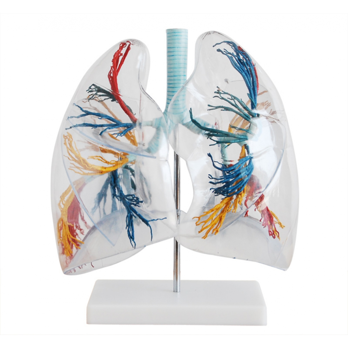
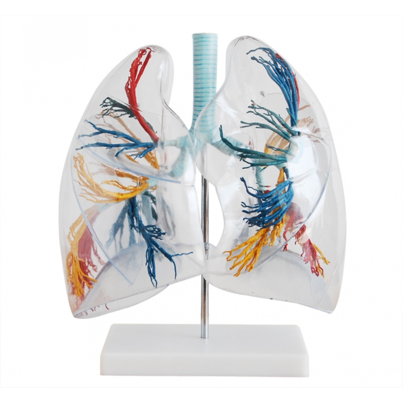
In respiratory anatomy, clarity is everything. The Transparent Lungs Model by MYASKRO delivers that clarity—literally. Encased in a crystal-clear outer shell, this anatomical model showcases the full bronchial tree, left and right lung structures, and the detailed anatomy of the hilus of the lung with unmatched visual precision. It’s a powerful tool for students, educators, and clinicians who want to see not just the lungs, but the entire pulmonary system in action.
Designed for high-impact learning and clinical relevance, this model brings the complexity of the respiratory tree into focus. Every primary, secondary, and tertiary bronchus is represented in full color, with surrounding vascular structures highlighted to emphasize the lung’s integration into the cardiovascular system. Learners can clearly identify the pulmonary arteries (blue), veins (red), and bronchi (yellow and blue), along with branching nerve pathways and the anatomical positioning of the tracheobronchial tree.
This model is more than just a teaching tool—it’s a visual revelation. The transparent casing allows students to view internal branching from all angles, reinforcing spatial understanding and three-dimensional orientation. Whether you're teaching airway structure in an anatomy lab, demonstrating pulmonary flow in a lecture hall, or training students in bronchoscopy navigation, this model provides the perspective needed to bridge concept with comprehension.
Perfect for:
As one respiratory instructor put it, “This is the only model where you can see everything that matters—bronchi, vessels, hilum—without having to open anything up.”
Mounted on a stable display base, the model is lightweight, portable, and crafted from high-durability materials. It’s ideal for repeated classroom use or one-on-one clinical demonstrations, offering years of value and visual precision.
For any educator looking to elevate how students see and understand the lungs, the MYASKRO Transparent Lungs Model is a clear choice—inside and out.
The Transparent Lungs Model by MYASKRO provides a rare advantage in anatomy education—complete visibility into one of the most intricate systems in the human body. From the surface contours to the deepest tertiary bronchi, this model lays out the entire pulmonary map in a way that is intuitive, engaging, and incredibly effective for teaching.
Encased in a durable, transparent shell, the lungs are fully visible from all sides, allowing learners to observe the branching architecture without obstruction. Every component is color-coded to differentiate critical structures:
This makes it easy for students to trace the flow of oxygen from the trachea through the bronchi and into the smaller airways, while also understanding how oxygenated and deoxygenated blood travel to and from the lungs. The inclusion of the hilus of the lung—the gateway where bronchi, arteries, veins, and nerves enter and exit—is a major highlight, helping learners visualize the lung’s role as both a respiratory and vascular organ.
The model supports a wide range of learning applications:
Its compact design allows for easy transport between classrooms or patient rooms, while the stable white base provides secure upright display. Made with medical-grade acrylic and non-toxic internal components, the model is built for durability and repeated handling without losing color or structural integrity.
As one anatomy lab director noted, “You can teach lung anatomy with a book—or you can show them this, and they’ll never forget it.”
Whether you're running a lecture, leading a skills lab, or guiding a clinical explanation, this model turns abstract lung diagrams into a living, breathing educational experience—visible in full, from every angle.
Understanding the lungs means understanding life at its most fundamental level—how we breathe, how oxygen reaches our cells, how the body sustains itself. The Transparent Lungs Model by MYASKRO gives students and educators the ability to explore that system with absolute clarity. No guesswork. No abstraction. Just visible, traceable anatomy from trachea to terminal branches.
Unlike traditional models that require disassembly or guesswork to explore the deeper structures, this model lays everything out in one intuitive display. You can rotate it, examine it from every angle, and explain the role of each part in context. This transforms your teaching from static instruction to immersive learning—especially when paired with real-world clinical examples like pneumonia, pulmonary embolism, or bronchial obstruction.
Its greatest strength lies in its ability to connect form with function. As students visually track the bronchial branches, they begin to understand how air is distributed across lung segments. As they examine the hilus and trace the entry and exit points of vessels and nerves, they see the lungs not as isolated organs, but as integrated hubs of respiration and circulation.
Educators across medical schools, respiratory therapy programs, and allied health institutions rely on this model to:
Patients, too, benefit from this visual approach. For those facing lung surgery, chronic pulmonary conditions, or post-operative recovery, this model helps them visualize what’s happening inside their own body. It fosters better communication, lowers anxiety, and empowers patient participation in their care decisions.
Crafted for clarity, built for daily use, and engineered to inspire understanding, the MYASKRO Transparent Lungs Model stands apart in its ability to make anatomy not just seen—but understood. It doesn’t just show structure—it shows connection, function, and life.
As one faculty member remarked, “With this model, students don’t just remember lung anatomy—they finally get it.”
And once they do, the knowledge stays with them—not just for exams, but for every patient they’ll one day care for.
Total Reviews (1)