
")
₹ 6,299 ₹ 22,379

₹ 8,599 ₹ 17,423

₹ 15,499 ₹ 22,379

₹ 3,599 ₹ 22,379

₹ 2,599 ₹ 10,579

₹ 5,899 ₹ 22,379
 – Myaskro")
₹ 3,899 ₹ 11,641

₹ 8,799 ₹ 25,895

₹ 4,499 ₹ 6,071

₹ 6,899 ₹ 17,530
 Premium Quality - MYASKRO")
₹ 1,599 ₹ 4,906

₹ 5,199 ₹ 7,290
 Dissectible Into 3-Parts - Myaskro")
₹ 2,599 ₹ 7,782

₹ 1,999 ₹ 4,550

₹ 1,899 ₹ 5,488
![Human Torso Model (Unisex) 23 Parts [85cm Tall] - MYASKRO](https://theanatomyshop.com/image/cache/catalog/Product%20Images/human%20torso%20model%2085cm%20unisex%20XC%2001-10x10.jpg "Human Torso Model (Unisex) 23 Parts [85cm Tall] - MYASKRO")
₹ 19,899 ₹ 38,757

₹ 2,699 ₹ 9,268

₹ 35,799 ₹ 57,779

₹ 10,299 ₹ 22,379

₹ 12,499 ₹ 34,179

₹ 3,499 ₹ 17,541

₹ 8,999 ₹ 12,865
 - Myaskro")
₹ 3,199 ₹ 4,865

₹ 4,399 ₹ 8,965
 | Myaskro")
₹ 18,299 ₹ 28,945
")
₹ 169,299 ₹ 184,965
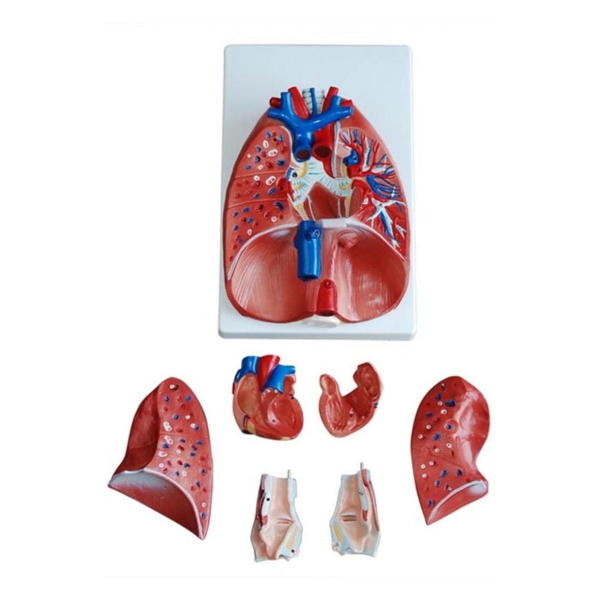
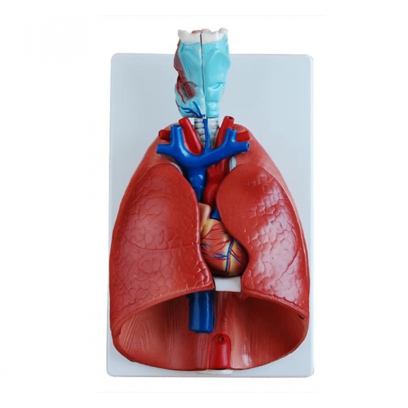
When it comes to teaching human anatomy, integration matters. The MYASKRO Larynx, Heart and Lungs Model combines three essential systems—respiratory, cardiovascular, and upper airway—into one highly detailed and interactive learning tool. Designed for educators and students who demand clarity, depth, and anatomical precision, this model offers a complete thoracic overview, ideal for building core knowledge and clinical confidence.
Crafted with vibrant color-coded structures and anatomically accurate contours, this model helps learners visualize the spatial relationships between the larynx, heart, lungs, trachea, bronchi, and diaphragm. It shows not only where these organs sit, but how they work together to power respiration, oxygen transport, and blood circulation.
Perfect for anatomy classes, nursing labs, OSCE prep, and patient education, the model can be used in both beginner and advanced training environments. It’s also an invaluable tool for respiratory therapy instructors teaching airflow, voice function, and pulmonary health from a systems-based approach.
Key components include:
Medical students use this model to learn the positioning and function of thoracic organs in situ. Nursing students reference it during respiratory assessment training. And instructors rely on it to visually explain complex interdependent processes—like gas exchange, voice production, and cardiac output—all in one integrated framework.
As one anatomy professor put it, “This is the model I reach for when I want students to connect the dots between breathing, circulation, and oxygenation.”
Mounted on a sturdy base and built from durable, non-toxic materials, this model stands up to daily classroom use while offering exceptional educational value. Whether it’s for demonstration or dissection, this tool brings together everything students need to understand the thoracic cavity—from larynx to lungs to heart—with clarity and impact.
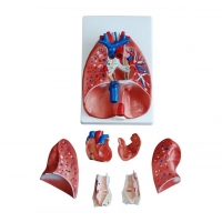
The MYASKRO Larynx, Heart and Lungs Model is more than just a visual aid—it's a dynamic, hands-on teaching tool that helps students engage with the full complexity of the thoracic cavity. Each part is carefully constructed to offer a detailed, layered understanding of the respiratory and cardiovascular systems, as well as their anatomical relationships and physiological interactions.
One of the most powerful features of this model is its modular design. It includes detachable lungs, heart components, and upper airway sections, allowing instructors and learners to break down the system piece by piece. Students can remove each lung to examine the bronchial tree and alveolar structures, explore the internal chambers of the heart, or isolate the larynx to understand vocal cord anatomy and its connection to respiration and phonation.
The model is color-coded for arteries (red), veins (blue), and air passages (light blue and white), making it easy to follow the path of blood and air throughout the body. This helps learners understand how oxygen enters the lungs, diffuses into the bloodstream, and is transported to organs via the heart—all while providing a clear visual of the anatomical route each process follows.
This comprehensive model is ideal for teaching:
Medical students benefit from the tactile experience of physically separating and examining components. It supports their understanding of system integration and reinforces procedural concepts such as chest assessments, cardiac auscultation points, or endotracheal tube placement. For nursing and paramedic training, it’s a go-to tool for teaching vital sign interpretation, airway management, and emergency anatomy.
The model’s durable construction and stable base make it suitable for repeated classroom use, lab demonstrations, and small group instruction. Its clean finish and detachable parts encourage curiosity and interaction—turning passive observation into active learning.
As one clinical educator shared, “This model makes complex systems feel intuitive. Students stop memorizing and start visualizing.”
With every component visible, removable, and anatomically accurate, the MYASKRO model delivers an unparalleled educational experience—designed to help learners build knowledge from the inside out.
In healthcare education, the ability to visualize systems holistically—not just in isolation—is essential. The MYASKRO Larynx, Heart and Lungs Model delivers that integrative learning experience with stunning anatomical accuracy and hands-on flexibility. Whether you're teaching first-year anatomy or preparing clinical students for real-world diagnostics, this model makes complex physiology visible, tangible, and unforgettable.
Its multi-part, removable structure empowers students to not just observe anatomy but to interact with it. They can dissect the model, trace airflow from the larynx through the bronchial tree, open the heart to explore its chambers, and examine the vascular networks that oxygenate the body. Each of these layers reinforces the vital connections between form and function, making it easier to retain knowledge and apply it with confidence during assessments or patient care scenarios.
Educators appreciate the model’s utility across disciplines. It fits seamlessly into anatomy courses, physiology labs, cardiopulmonary units, and OSCE prep. Nursing faculty use it to support respiratory and cardiovascular instruction. Medical schools employ it for system-based learning. Allied health programs include it in airway and circulatory assessment training. And because the model is clear, clean, and approachable, it’s even ideal for patient education and community health workshops.
Its compact, vertical mount makes it a standout piece in any classroom or simulation environment, offering both portability and presence. Built with non-toxic, classroom-grade resin, the model holds up under daily use while maintaining its vibrant detail and structure. It’s as durable as it is educational—built for years of repeat instruction.
Perhaps most importantly, the MYASKRO model gives learners the ability to connect the dots between systems. They no longer see the lungs, heart, and larynx as isolated topics, but as co-dependent players in the critical processes of oxygenation, circulation, and respiration. That shift—from memorization to integration—is where deep understanding begins.
As one medical instructor put it, “This model bridges everything we teach in first-year anatomy. It doesn’t just show the parts—it shows how they work together.”
With the MYASKRO Larynx, Heart and Lungs Model, you’re not just teaching organ systems—you’re building system thinkers. Learners who can see, understand, and apply what they know with clarity and purpose.
Total Reviews (0)