


₹ 1,599 ₹ 4,537
")
₹ 1,699 ₹ 4,561
 With Ligaments")
₹ 1,699 ₹ 4,955

₹ 1,699 ₹ 4,955

₹ 1,699 ₹ 4,955

₹ 8,499 ₹ 25,919

₹ 5,799 ₹ 15,157

₹ 1,399 ₹ 5,427
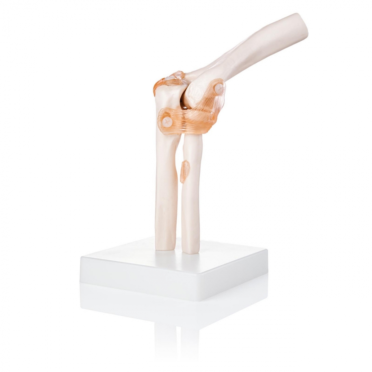
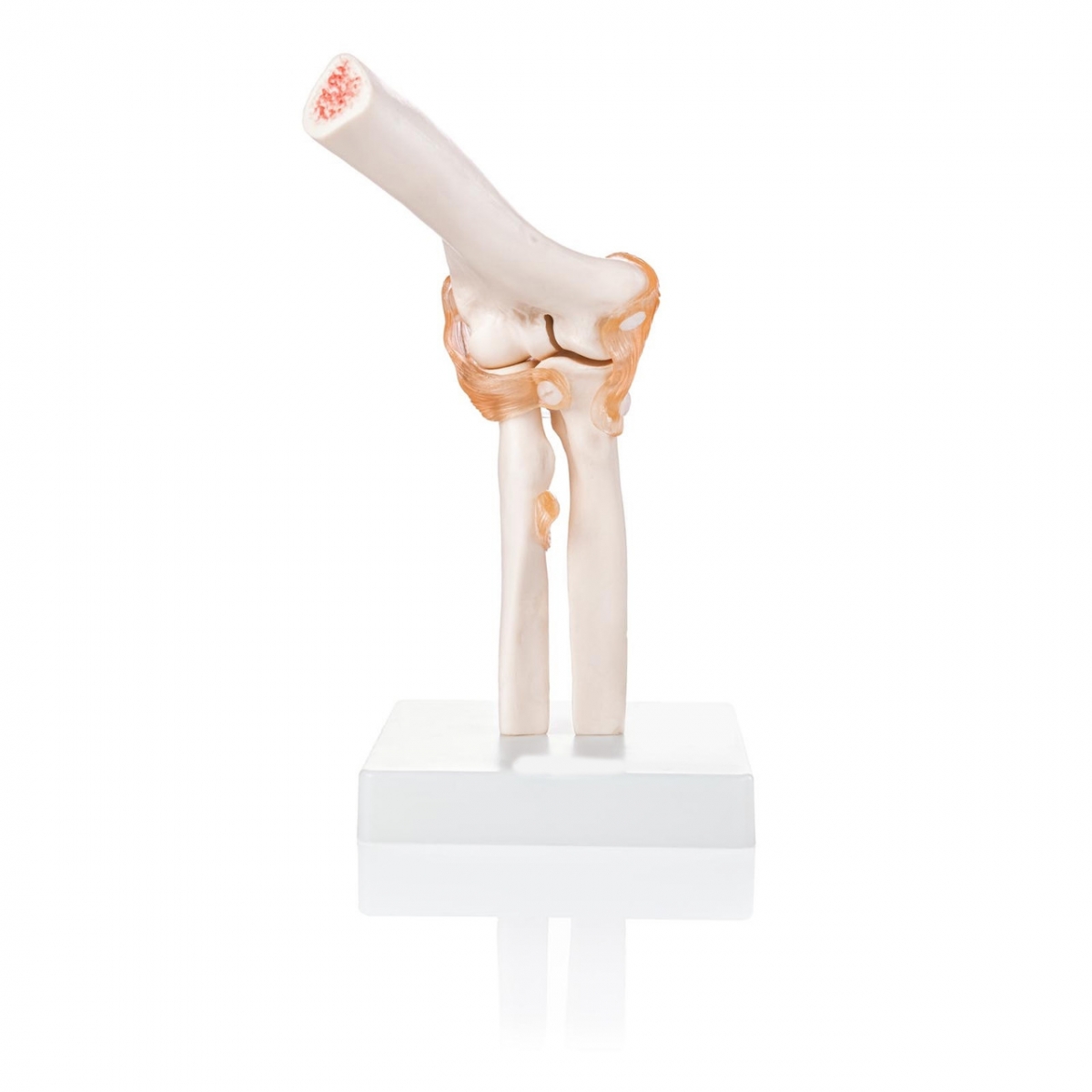
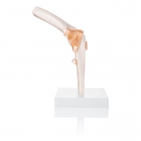
The human elbow is more than a hinge—it's a precisely coordinated articulation of bones, ligaments, and movement patterns. The MYASKRO Life-Size Elbow Joint Model with Ligaments brings this intricate structure to life with medical-grade realism, making it ideal for orthopedic teaching, physiotherapy, medical education, and patient consultations.
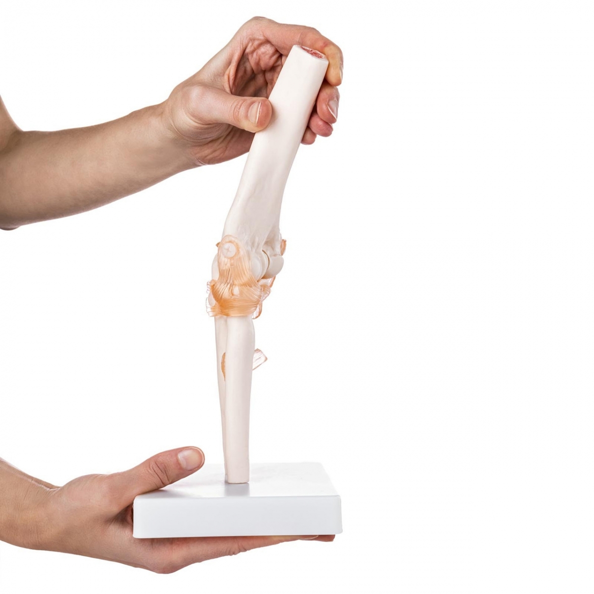
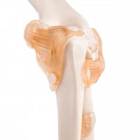
This model features the full articulation of the humerus, radius, and ulna, joined with semi-flexible ligaments to demonstrate real movement. The ligaments aren’t just decorative—they’re functional, allowing clear demonstration of flexion, extension, and rotational movements like pronation and supination. Whether you're a medical student learning surface anatomy or a professional explaining joint dysfunction to a patient, this model delivers unmatched clarity.
Crafted from non-toxic, high-strength PVC, the model is safe, durable, and lightweight. It comes mounted on a sturdy base, yet the joint itself is fully manipulable—ideal for table-side teaching or in-class instruction. This makes it a preferred tool for orthopedic surgeons, chiropractors, physiotherapists, sports trainers, and rehabilitation experts.
A common query: “Does it replicate the natural range of motion?” The answer is yes. Thanks to its functional ligaments and accurate alignment, the model allows realistic elbow articulation, making it suitable for demonstrating injury mechanisms, rehab protocols, and anatomical relationships.
Searches like “functional elbow joint model,” “elbow model with ligaments,” “life-size elbow joint anatomy,” and “PVC elbow joint teaching tool” all lead here—for good reason. This model delivers what those terms promise: function, detail, and durability.
“I use this daily during rounds at my ortho clinic. My interns and patients both understand conditions like ligament tears and arthritis much faster when I show them this,” says Dr. Vivek Rana, Orthopedic Consultant, Max Hospital, Dehradun.
When learning, teaching, or treating conditions of the elbow, this model becomes more than just a visual—it becomes a bridge between anatomy and application. Built to last. Designed to move. Ready to educate.
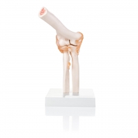
The MYASKRO Elbow Joint Model isn’t just a visual display—it’s a dynamic, functional model built for hands-on anatomical exploration. Whether you’re explaining joint movements to a classroom or helping a patient understand the root of their discomfort, this life-size replica offers a high-fidelity experience that enhances both comprehension and communication.
The model includes all major anatomical structures of the elbow:
Thanks to its flexible ligament construction, this model enables demonstration of key joint actions:
Frequently used in orthopedic teaching modules, physiotherapy sessions, OSCE prep, and joint assessment tutorials, this model brings theory to life. Trainers can clearly illustrate conditions like tennis elbow, medial ligament strain, or post-surgical range limitation—while students get a tactile feel for bone positioning and ligament tension.
Made from high-density, non-toxic PVC plastic, the model is resistant to wear and ideal for repetitive motion. It is mounted on a white display base that ensures stability during use. The base can be detached, allowing for handheld demonstration or close-up table work.
It aligns well with search queries such as “elbow joint ligament model,” “life-size anatomical elbow with soft tissue,” “PVC elbow demonstration model,” and “clinical elbow training tool.” And every one of those use-cases is fully supported by the design and performance of this model.
“Our batch used it for practical anatomy sessions and it made identifying the radial head and ligament attachments so much easier,” says Reetika Sharma, Final Year MBBS, IGMC Shimla.
Compact, durable, and clinically focused, this elbow model is built for education in motion—because learning anatomy should never be static.
Understanding joint anatomy isn’t just about memorizing parts—it’s about grasping how those parts move, interact, and respond to stress. The MYASKRO Life-Size Elbow Joint Model with Ligaments delivers exactly that: a full sensory learning experience where bones, ligaments, and movement all come together in perfect anatomical harmony.
From the moment you place it on your desk or clinic counter, this model invites interaction. Medical students explore the trochlea and olecranon groove. Physiotherapists use it to illustrate restricted pronation. Orthopedic surgeons break down injury patterns for pre-surgical briefings. It adapts to the user and elevates the lesson, no matter who’s holding it.
Because the ligaments are not just static—they flex, shift, and demonstrate resistance—it’s ideal for explaining real-life elbow mechanics and common pathologies. Whether you’re discussing chronic epicondylitis, instability following trauma, or post-operative joint care, this model strengthens your case with something visual and concrete.
Users frequently ask: “Will this model last in daily use?” It will. The materials—non-toxic PVC and reinforced polymer ligaments—are chosen for their strength and longevity. Designed for movement, resistant to wear, and mounted securely, this model thrives under frequent use in demanding academic or clinical settings.
It aligns with key semantic searches like “elbow model with soft ligaments,” “functional joint model PVC,” “orthopedic demo elbow tool,” and “anatomy joint model for physiotherapy.” Because it checks every box a user might look for: scale, motion, durability, and precision.
“I use this in our sports rehab department every week. It helps patients understand their joint limitations and rehabilitation goals instantly,” shares Dr. Aayush Mehra, Sports Physiotherapist, DY Patil Hospital, Mumbai.
In teaching and healing alike, clarity leads to confidence. And confidence leads to better outcomes. This elbow joint model doesn’t just explain anatomy—it empowers it. With every movement it demonstrates, it helps someone better understand their body, their injury, or their future.
Total Reviews (0)