

Anatomic Details
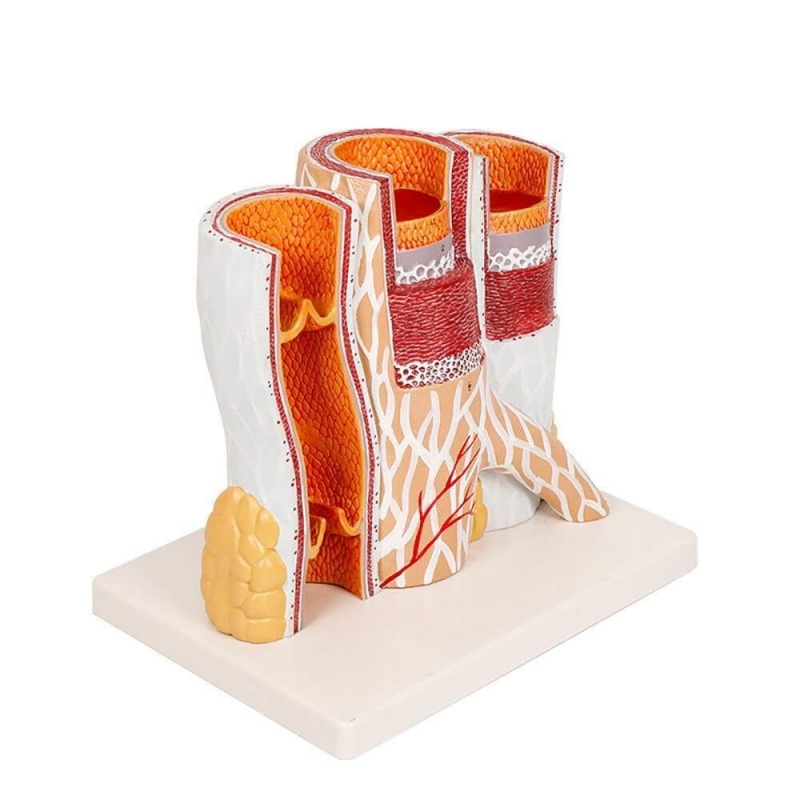
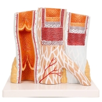
Giant longitudinal cross-sections of a muscular artery and a medium vein.
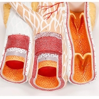
Demonstrates tunica intima (endothelium, subendothelial layer, internal elastic lamina), tunica media (circular smooth muscle; external elastic lamina in artery), and tunica adventitia (collagen, vasa vasorum, nervi vasorum).
Vein segment with bicusp venous valves (leaflets, sinus, commissures).
Central block showing arteriole → capillary plexus → venule (microcirculation).
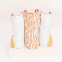
Surrounding adipose tissue and peri-vascular connective tissue.
Learning objectives
Differentiate artery vs vein wall architecture and lumen morphology; explain pulsatile conduit (artery) vs capacitance vessel (vein).
Trace flow through arterioles–capillaries–venules and relate to diffusion/exchange.
Correlate structure with common pathology: atherosclerosis/intimal plaque, aneurysm (media weakening), varicose veins/valvular incompetence, DVT sites, phlebitis.
Use for OSCE/viva: identify layers, elastic laminae, venous valve components, and vasa vasorum.
Specifications
Approx. 27 × 20 × 24.5 cm on a stable base (classroom display size).
Material: Medical Grade PVC.
Cleaning: mild detergent or 70% alcohol wipes; avoid solvents/heat.
Intended use: instructional model for physiology, pathology, cardiology, nursing, and patient education.
Total Reviews (0)