


₹ 7,999 ₹ 22,379
 With Hard Backing, Aluminium Frame & Hanging Hooks")
₹ 2,800 ₹ 4,561
 With Special Lamination")
₹ 1,599 ₹ 3,865
 With Ligaments")
₹ 1,699 ₹ 4,955

₹ 20,599 ₹ 28,965

₹ 31,699 ₹ 42,645

₹ 11,799 ₹ 18,945
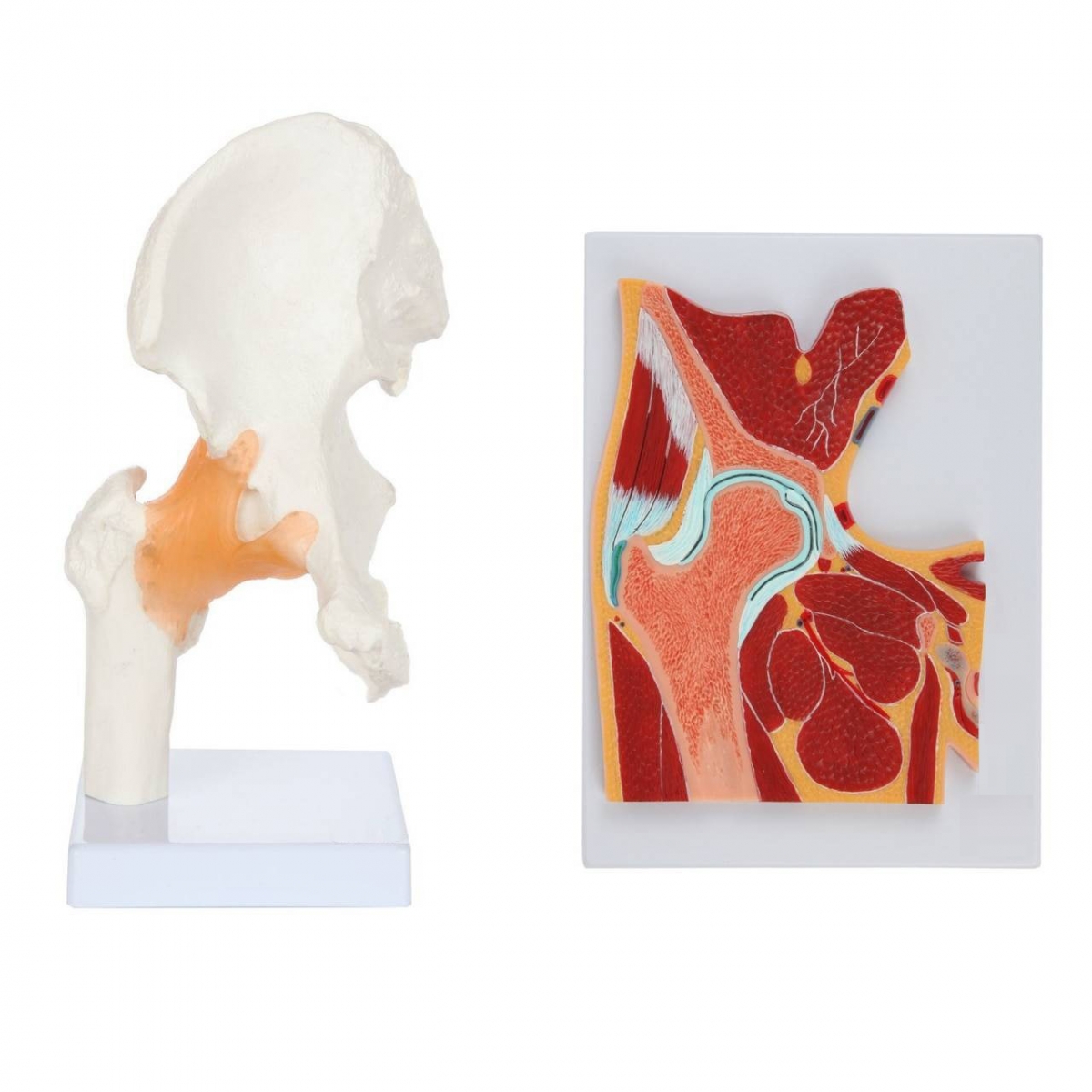
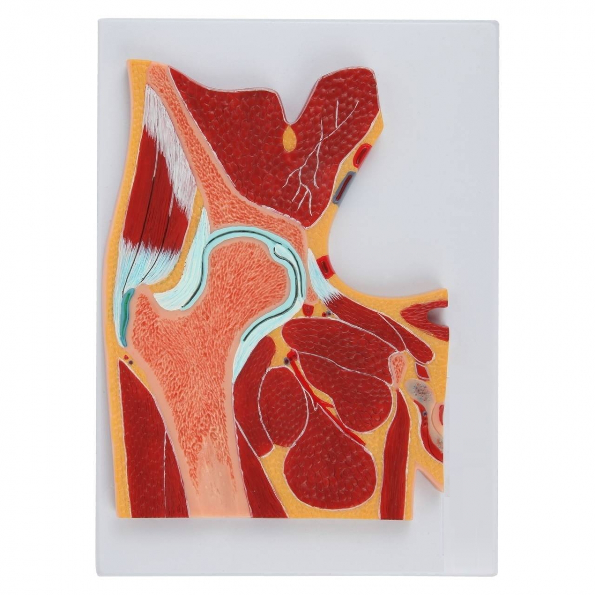
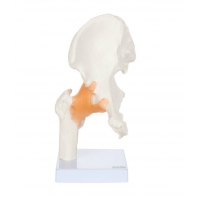
MYASKRO Hip Joint & Cross-Section Anatomy Model Set
A dual-view teaching set that pairs a life-size hip joint with an enlarged cross-section of the lateral hip. Ideal for MBBS/BDS foundations, PT/OT training, OSCE/OSPE stations, and skill-lab demonstrations.
Key Benefits
Two perspectives, one set: Articulated femoral head–acetabulum model + large relief cross-section for 3D and planar learning.
Clinically accurate landmarks: Ilium/ischium/pubis, acetabular rim and labrum, hyaline articular cartilage, femoral head–neck, greater trochanter, joint capsule.
Myofascial context: Shows gluteal group, iliopsoas and adductor planes with neurovascular pathways for approach planning (posterior/anterolateral).
Procedure mapping: Demonstrate hip biomechanics, capsular tightness, trochanteric pain syndrome discussion, and safe surface anatomy for injections (education only).
Built for classrooms: Rigid, wipe-clean surfaces; colour-coded layers for fast recall; stable base for bench teaching.
Teach the hip from bone to soft tissue. A life-size joint model clarifies articulation and movement, while a high-contrast cross-section reveals muscles, capsule and labrum—perfect for lectures, tutorials and exams.
This MYASKRO set integrates macro 3D anatomy with a sectional view so learners can correlate bony landmarks to soft-tissue planes. The joint component highlights the femoral head seated in the acetabulum with articular cartilage and the acetabular labrum, useful for explaining stability, load transfer and degenerative change. The cross-section maps the capsule, surrounding musculature (gluteals, iliopsoas, adductors), and vascular/nerve corridors—supporting discussions on surgical approaches, bursitis, femoroacetabular impingement and osteoarthritis. Durable, classroom-ready construction withstands daily handling and routine cleaning.
Teaching Objectives & Skills Practised
Identify hip osteology and key surface landmarks (ASIS, greater trochanter, acetabular rim).
Describe capsule, labrum and articular cartilage roles in stability and range of motion.
Correlate cross-sectional planes with imaging (X-ray/MRI basics) and clinical approaches.
Explain common pathologies: OA, FAI, capsular tightness, trochanteric pain syndrome.
Plan OSCE/OSPE demonstrations for hip examination and movement testing.
What’s Included
1 × Life-size hip joint model (ilium–ischium–pubis with femoral head/neck; highlighted cartilage/labrum & capsule).
1 × Enlarged coloured cross-section of the hip region on display board.
Total Reviews (0)