

₹ 6,999 ₹ 14,711
 – Myaskro")
₹ 3,899 ₹ 11,641

₹ 3,599 ₹ 22,379

₹ 8,999 ₹ 12,865
 | Myaskro")
₹ 10,899 ₹ 18,945
Overview
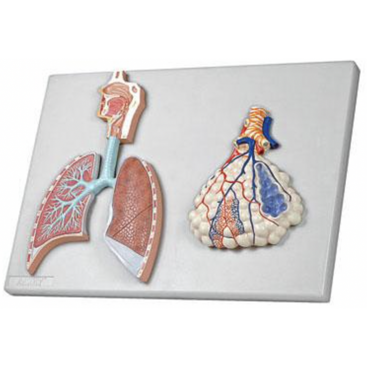
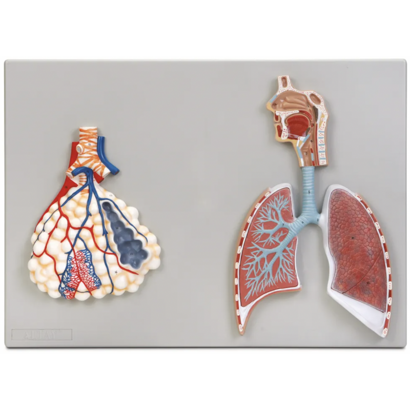
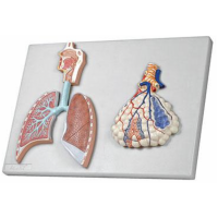
Two-piece teaching set illustrating the conducting airways to lung parenchyma and an enlarged acinus/alveolar sac. Designed for rapid correlation of structure with ventilation–perfusion and gas exchange. Instructional aid; not a medical device.
Components
Upper airway–lung panel: Nasal cavity → pharynx → larynx → trachea → main/lobar/segmental bronchi; cutaway lung parenchyma with bronchioles and terminal/respiratory bronchioles.
Enlarged alveolar unit: Alveolar ducts and sacs with inter-alveolar septa, dense pulmonary capillary network, and the respiratory membrane (type I pneumocyte, fused basement membrane, capillary endothelium) schematically indicated.
Learning objectives
Differentiate conducting vs respiratory zones and trace airflow from nares to alveoli.
Explain gas exchange across the alveolar–capillary barrier and principles of V/Q matching.
Discuss pathology at each level: bronchospasm/mucus plugging (asthma), consolidation (pneumonia), septal destruction (emphysema), interstitial/alveolar edema.
Support OSCE stations: airway mapping, lobar/segmental bronchus identification, diffusion concepts.
Specifications
Medical-Grade PVC relief models on stable backplates; high-contrast, hand-painted detail.
Wall-mount ready; wipe-clean (mild detergent or 70% alcohol; avoid solvents/heat).
Who it’s for
Medical and physiotherapy programs, nursing colleges, pulmonary rehab education, hospital skill labs, and patient counselling.
Total Reviews (0)