


 – Myaskro (USA) | Dual View with Study Guide")
₹ 8,999 ₹ 53,035
 With Special Rigid Lamination and Plastic Rollers")
₹ 1,599 ₹ 4,329

₹ 7,999 ₹ 22,379

₹ 17,699 ₹ 31,677

₹ 5,499 ₹ 10,319

₹ 40,899 ₹ 110,750

₹ 3,599 ₹ 22,379

₹ 5,899 ₹ 22,355
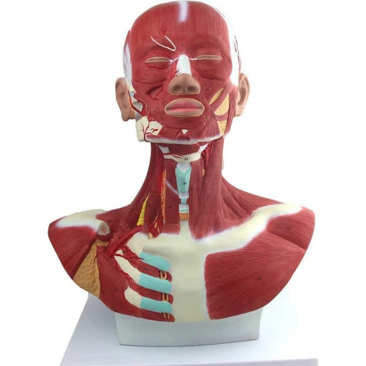
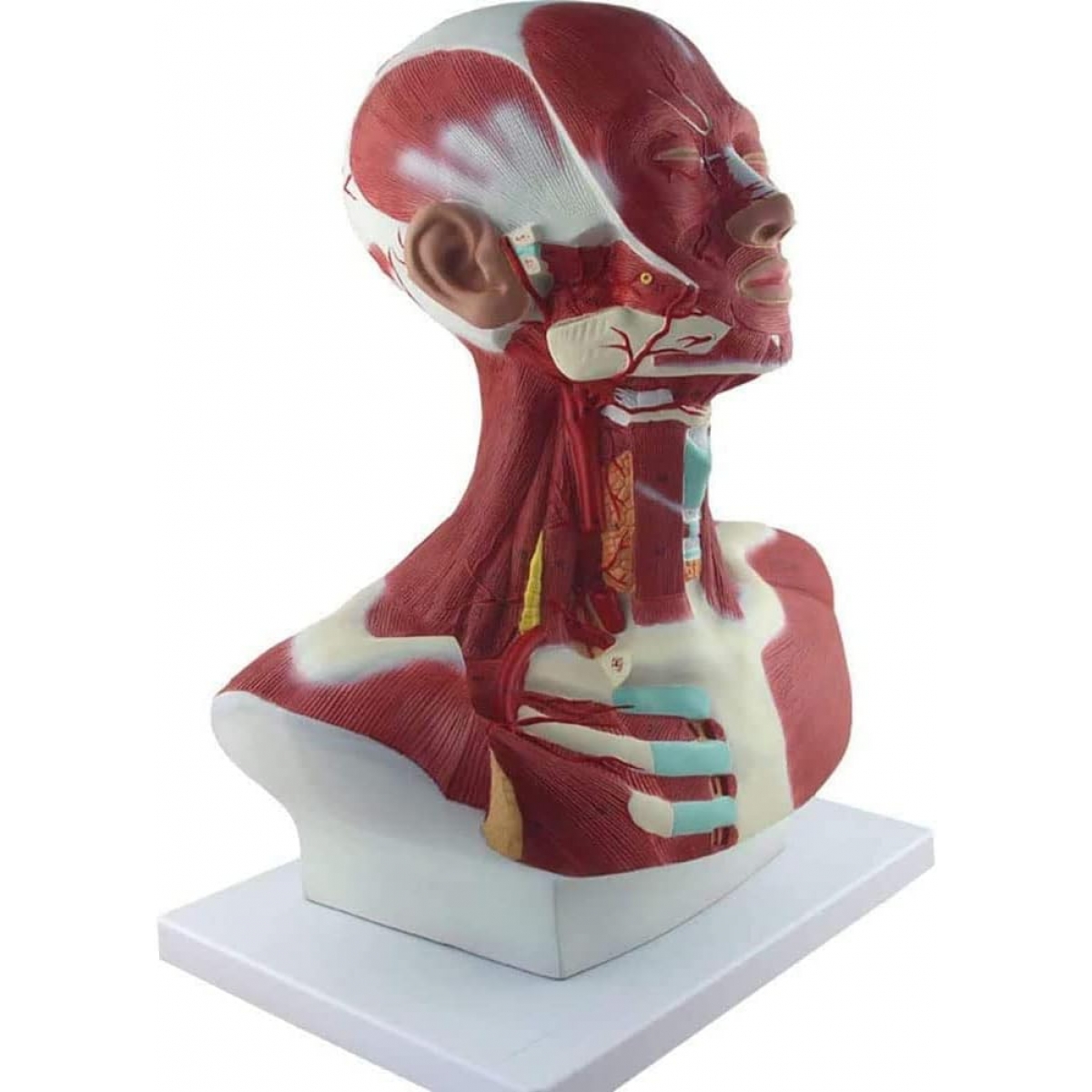
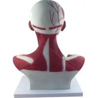
Head, Neck & Chest Muscle Anatomical Model (Life-size bust)
A clinically detailed teaching bust showing the musculature of the face, neck and upper thorax with key neurovascular and airway landmarks. Numbered relief sculpting makes it easy to identify structures at a glance—ideal for demonstrations, viva prep and OSCE stations.
What you’ll study (with brief lay cues):
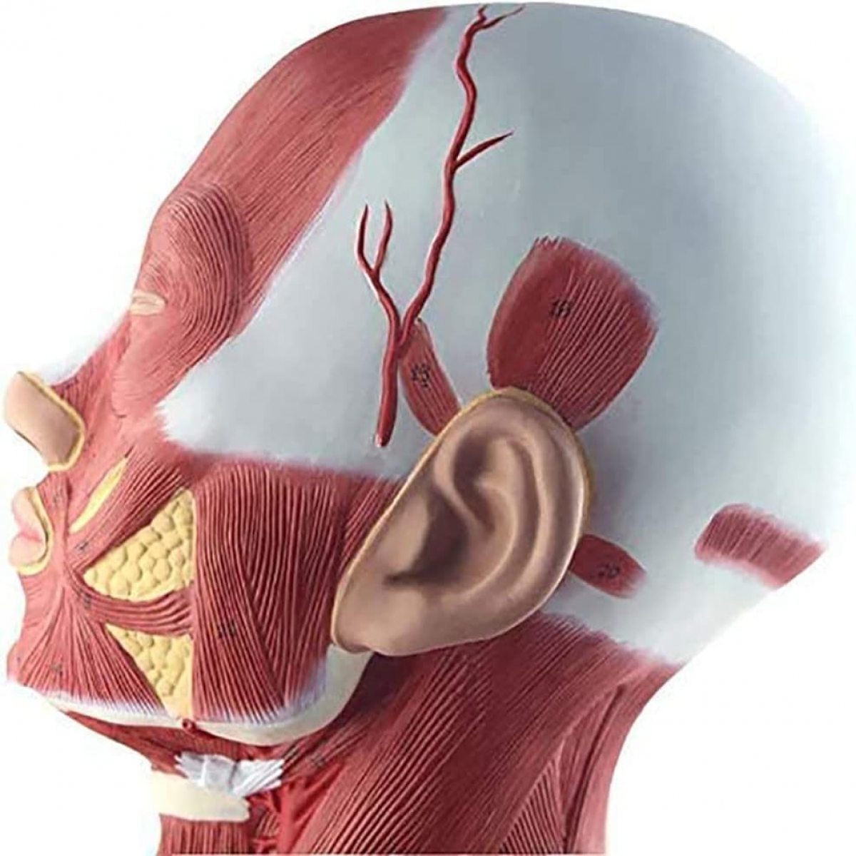
Muscles of facial expression—frontalis, orbicularis oculi/oris, buccinator, zygomaticus (facial movement & mimic).
Muscles of mastication—masseter, temporalis (jaw closing power).
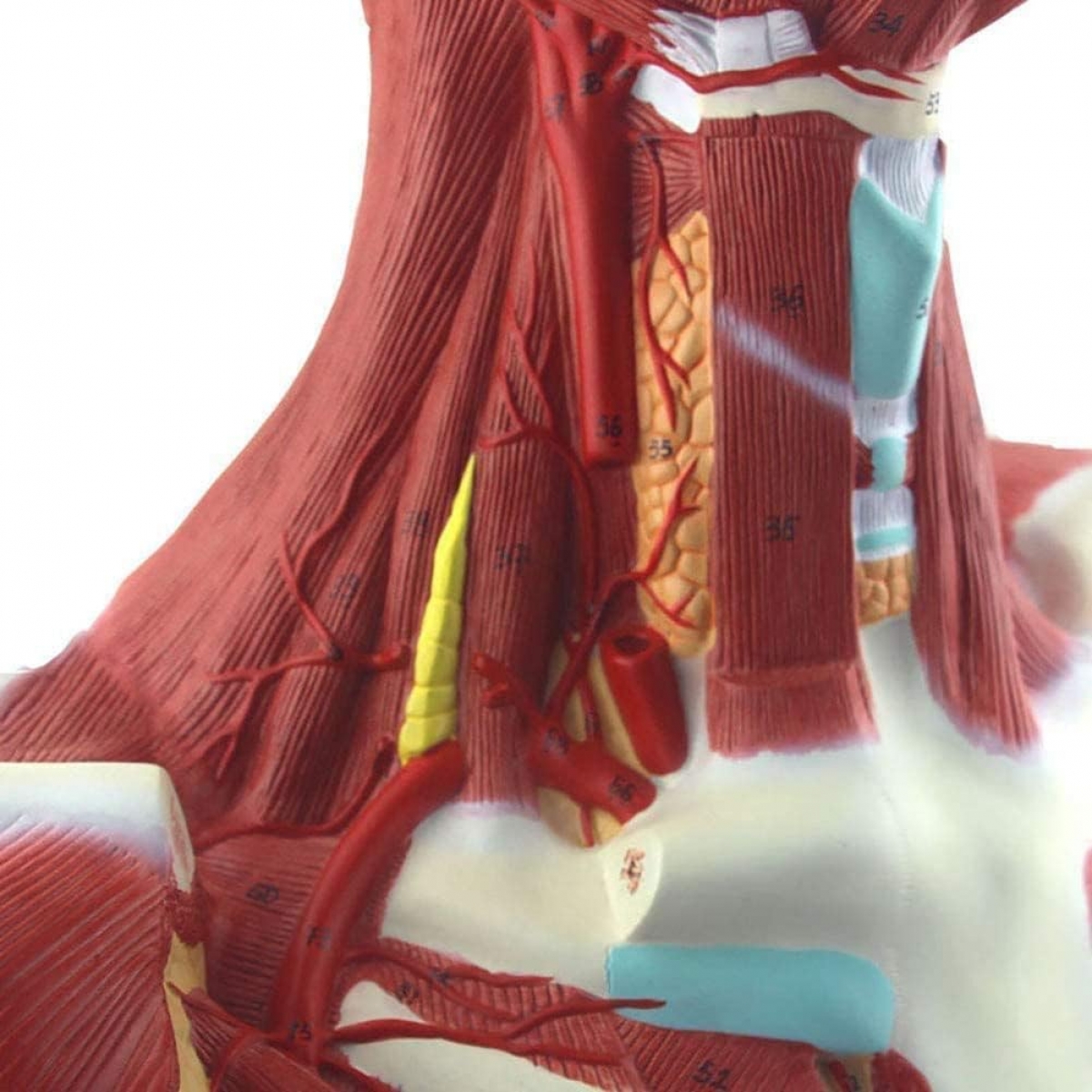
Cervical muscles—sternocleidomastoid, scalenes, infrahyoids incl. sternohyoid/omohyoid (neck posture, swallowing).
Posterior neck & shoulder—trapezius, upper pectoral region (scapular control, chest wall).
Airway & laryngeal cartilages—hyoid, thyroid & cricoid cartilages; tracheal rings (intubation/surgical airway landmarks).
Carotid sheath contents—common carotid artery, internal jugular vein (pulse, central line relevance).
Glands & fat pads—parotid region, buccal fat (procedural safety zones).
Costal cartilages 1–3 (chest wall continuity from neck to thorax).
Why educators choose this model
Near life-size 360° visualization for lecture halls and skill labs.
Numbered landmarks: simplifies quizzing and self-testing.
Durable, wipe-clean PVC with hand-painted detail; fixed on a stable base.
Perfect for MBBS/BDS, nursing, physiotherapy, ENT/anesthesia teaching, and simulation centers.
Total Reviews (0)