


₹ 2,199 ₹ 4,865

₹ 30,999 ₹ 47,845

₹ 15,499 ₹ 22,845

₹ 15,899 ₹ 29,353

₹ 33,499 ₹ 41,655

₹ 31,299 ₹ 45,955

₹ 10,899 ₹ 22,355
 With Special Lamination")
₹ 1,599 ₹ 3,865

₹ 1,599 ₹ 3,775

₹ 2,299 ₹ 6,921
 | Myaskro")
₹ 2,899 ₹ 5,753

₹ 1,999 ₹ 4,550

₹ 11,199 ₹ 17,865

₹ 9,999 ₹ 22,379

₹ 35,799 ₹ 57,779

₹ 41,999 ₹ 69,579

₹ 21,299 ₹ 45,979
 – Myaskro")
₹ 2,599 ₹ 5,465

₹ 64,499 ₹ 78,965
 | Myaskro")
₹ 22,299 ₹ 28,945
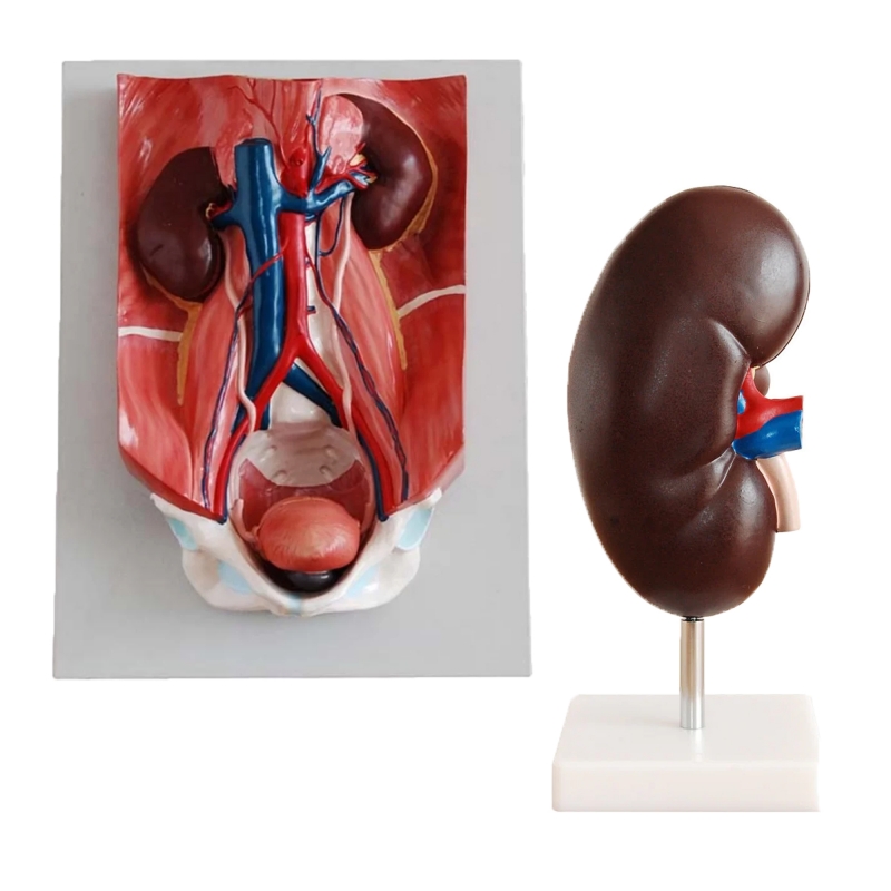
MYASKRO Urinary Anatomy Model Bundle
Dual-model teaching kit pairing a posterior abdominal wall–pelvis board (with kidneys and great vessels) and a life-size kidney model. Built for MBBS/BSc Nursing, physiology/anatomy labs, and OSCE/OSPE prep.
Key Benefits
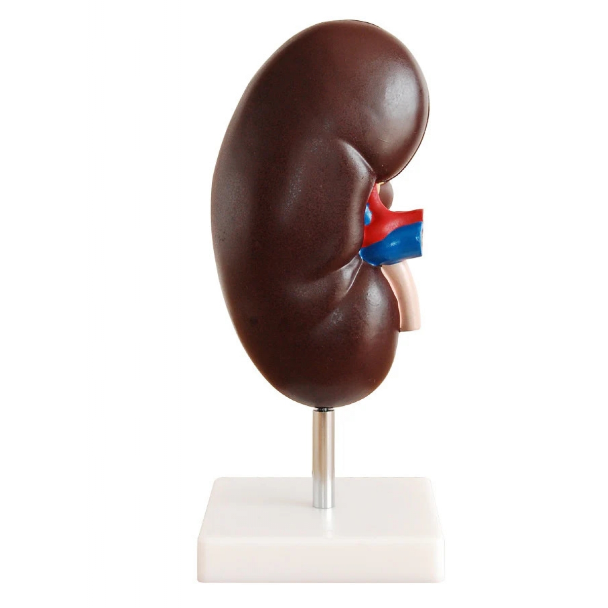
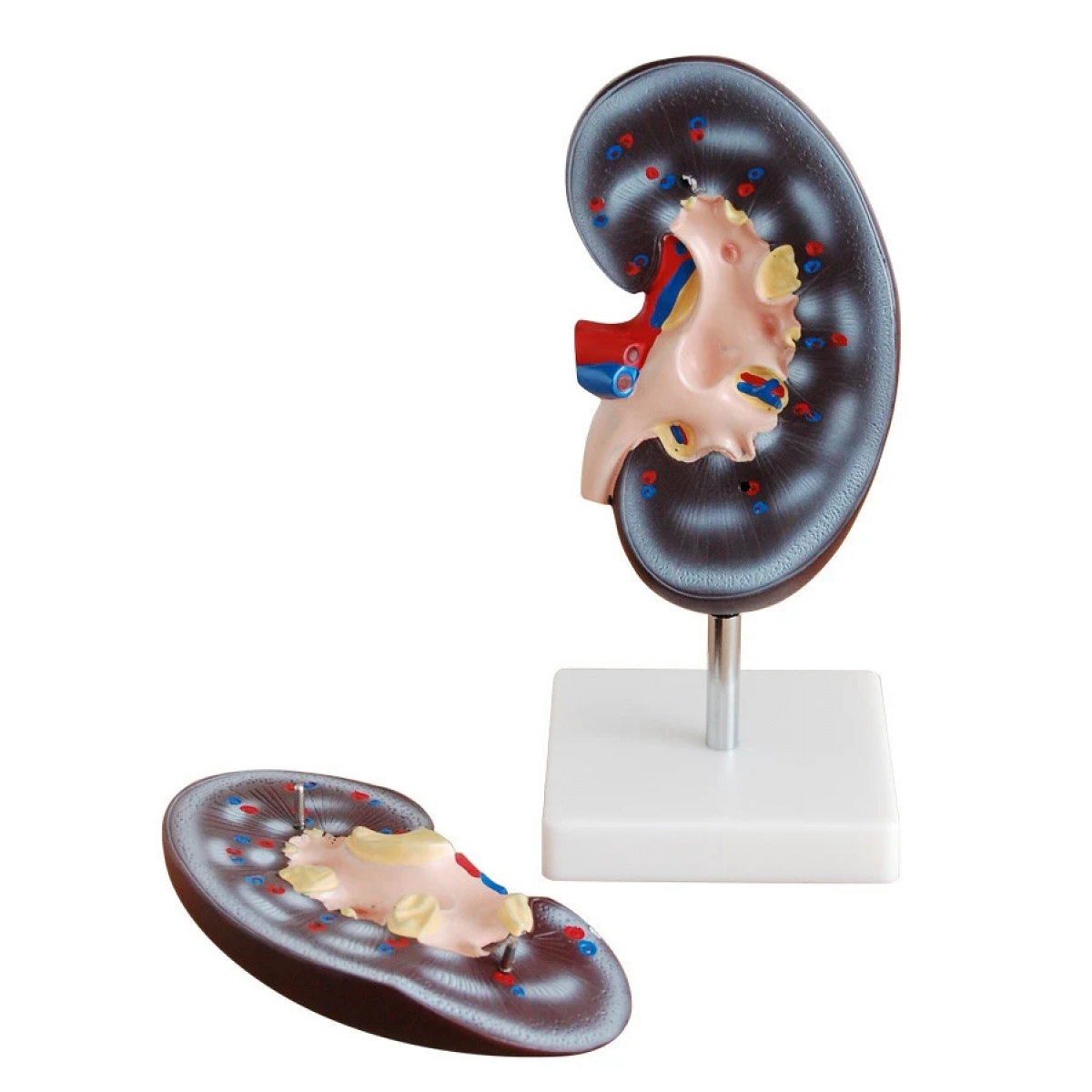
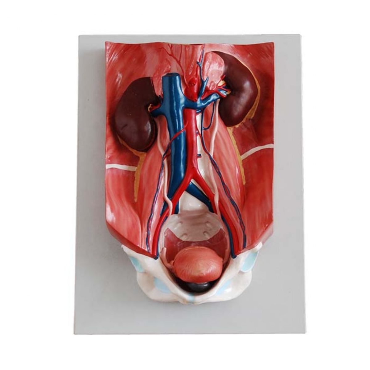
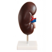
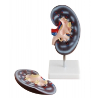
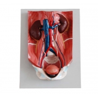
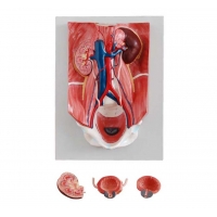
Two complementary views: Regional posterior abdominal wall with kidneys, ureters and bladder plus a stand-mounted kidney for focused study.
Clear spatial relations: Shows aorta/IVC and iliac vessels alongside kidneys, psoas region, ureters and pelvic inlet leading to the urinary bladder.
Renal hilum orientation: Identify renal artery, vein and ureter at the hilum; discuss cortex–medulla concept using the kidney model (theory).
Clinical correlation: Map obstruction sites (PUJ/UVJ), renal vascular relations and catheter-free bladder orientation for imaging basics (education only).
Classroom-ready: Life-size scale, high-contrast colours, wipe-clean surfaces and stable bases for repeated handling.
Teach urinary anatomy from kidneys to bladder: a posterior abdominal wall–pelvis board plus a life-size kidney model—ideal for colleges and skill labs.
This MYASKRO set links macro-regional and organ-level views of the urinary system. The wall board demonstrates both kidneys with their relations to the aorta and inferior vena cava, the course of the ureters across the pelvic brim, and the urinary bladder within the pelvis—useful for explaining vascular supply, ureteric constrictions and cross-sectional imaging orientation. The accompanying kidney model highlights external surfaces and the renal hilum (artery, vein, ureter) to reinforce anterior–posterior ordering and the concept of cortex/medulla (teaching). Durable construction makes the set ideal for lectures, vivas and OSCE stations.
Teaching Objectives & Skills Practised
Identify kidneys, ureters and urinary bladder with major vessels and muscular relations.
Describe renal hilum structures and their typical anterior–posterior arrangement.
Trace ureteric course and common narrowing points (PUJ, crossing iliac vessels, UVJ).
Correlate anatomy with CT/USG orientation and surface landmarks.
Build OSCE/OSPE stations integrating renal and pelvic urinary anatomy.
What’s Included
1 × Posterior Abdominal Wall–Pelvis Urinary Board (kidneys, aorta/IVC, ureters, bladder)
1 × Life-Size Kidney Model on Stand (external features with hilum)
Total Reviews (0)