
 With Special Lamination")
₹ 1,599 ₹ 3,865

₹ 1,599 ₹ 3,775

₹ 2,299 ₹ 6,921
 | Myaskro")
₹ 2,899 ₹ 5,753

₹ 1,999 ₹ 4,550

₹ 11,199 ₹ 17,865

₹ 9,999 ₹ 22,379

₹ 10,899 ₹ 22,355

₹ 13,799 ₹ 18,965

₹ 35,799 ₹ 57,779

₹ 41,999 ₹ 69,579

₹ 21,299 ₹ 45,979

₹ 64,499 ₹ 78,965

₹ 7,399 ₹ 12,865

₹ 18,999 ₹ 31,845
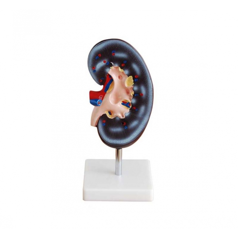
Kidney Model (Enlarged) – Myaskro
An enlarged, cut-section kidney for clear renal anatomy teaching. The one-piece model on a sturdy stand displays the cortex, medulla (renal pyramids & columns), papillae, minor/major calyces, renal pelvis, proximal ureter, and renal artery/vein with color cues. Great for bedside explanations, viva prep, and OSCEs.
Teach & explain (with quick lay cues):
Urine pathway—papilla → calyces → pelvis → ureter (how urine drains from the kidney).
Blood supply overview—arterial/venous entry at the hilum (why vascular disease affects filtration).
Common clinical points—obstruction sites, stone lodging, hydronephrosis basics.
Build & use
Enlarged single-piece model on stand—visible from across the room.
Durable, wipe-clean PVC with vivid, numbered landmarks.
Ideal for MBBS/BDS, nursing, urology/nephrology, physiotherapy and patient education.