


₹ 5,799 ₹ 7,756

₹ 5,299 ₹ 11,523
 With 7 Dissectible Parts - Myaskro®")
₹ 2,399 ₹ 7,262
 With Special Lamination")
₹ 1,599 ₹ 3,865

₹ 4,899 ₹ 21,199

₹ 21,599 ₹ 34,179

₹ 9,299 ₹ 16,865
")
₹ 169,299 ₹ 184,965
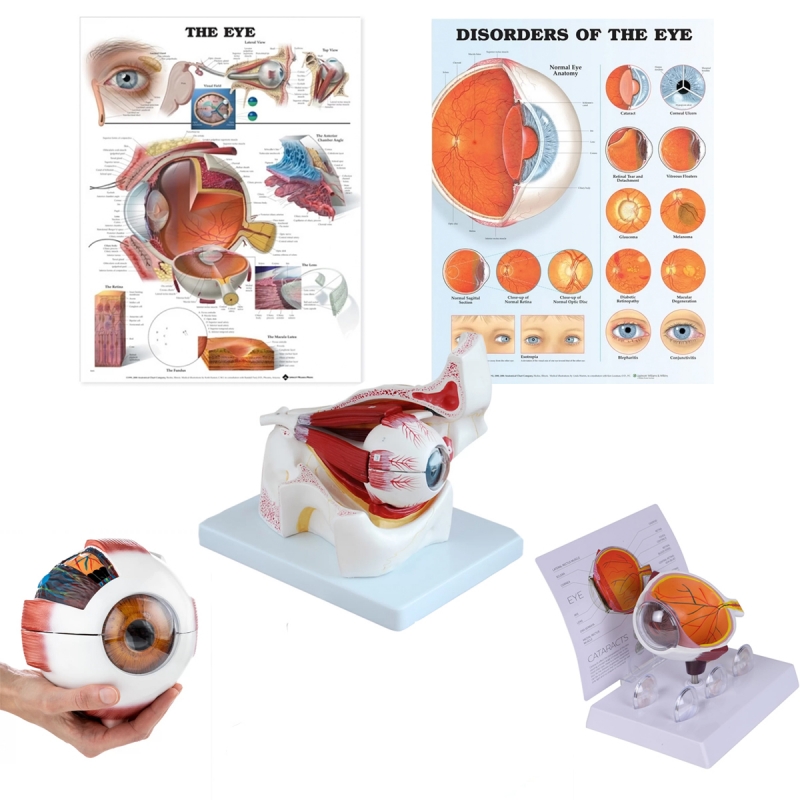
Components
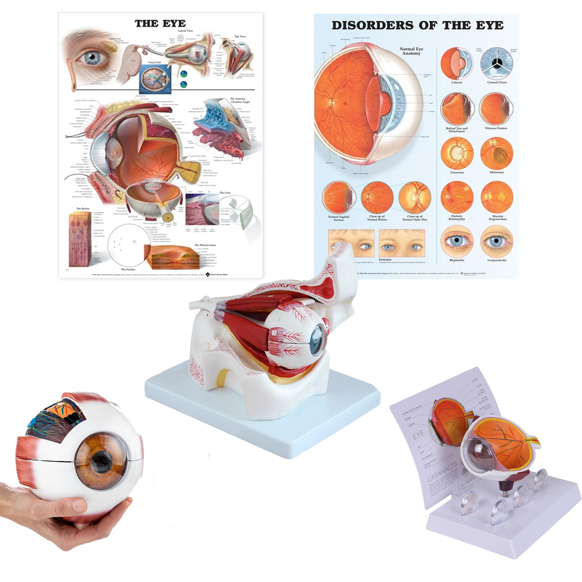
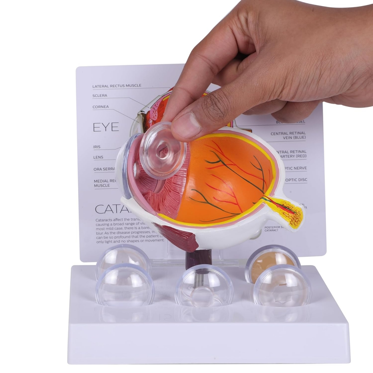
Cataract Eye Model 4 Times Enlarged with 5 Interchangeable Lenses: clear lens plus representative nuclear sclerosis, cortical spokes, posterior subcapsular opacity, and dense/mature cataract; demonstrates acuity loss and glare concepts.
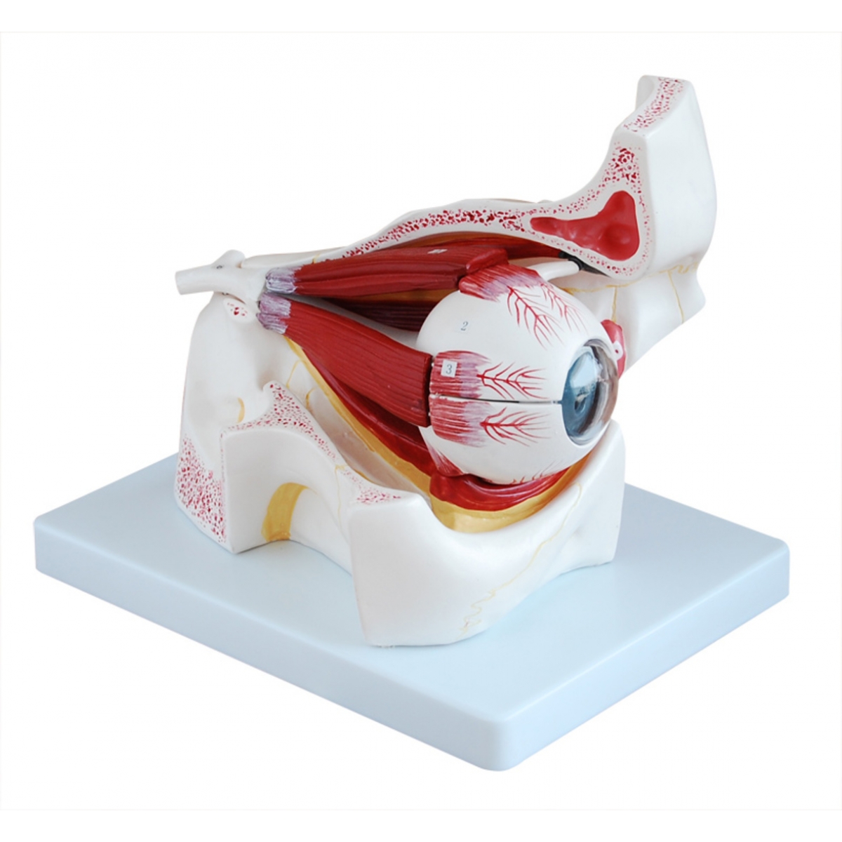
Eye with Orbit Model 3 Times Enlarged with 10 Removable Parts: Bony orbit with extraocular muscles (SR, IR, MR, LR, SO with trochlea, IO), lacrimal gland, optic nerve and canal; sectional apex/fissures.
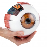
Eye Model 6 Times Enlarged with 6 Removable Parts: Sclera–choroid–retina shell, cornea, iris–ciliary body, crystalline lens, vitreous body, optic disc.
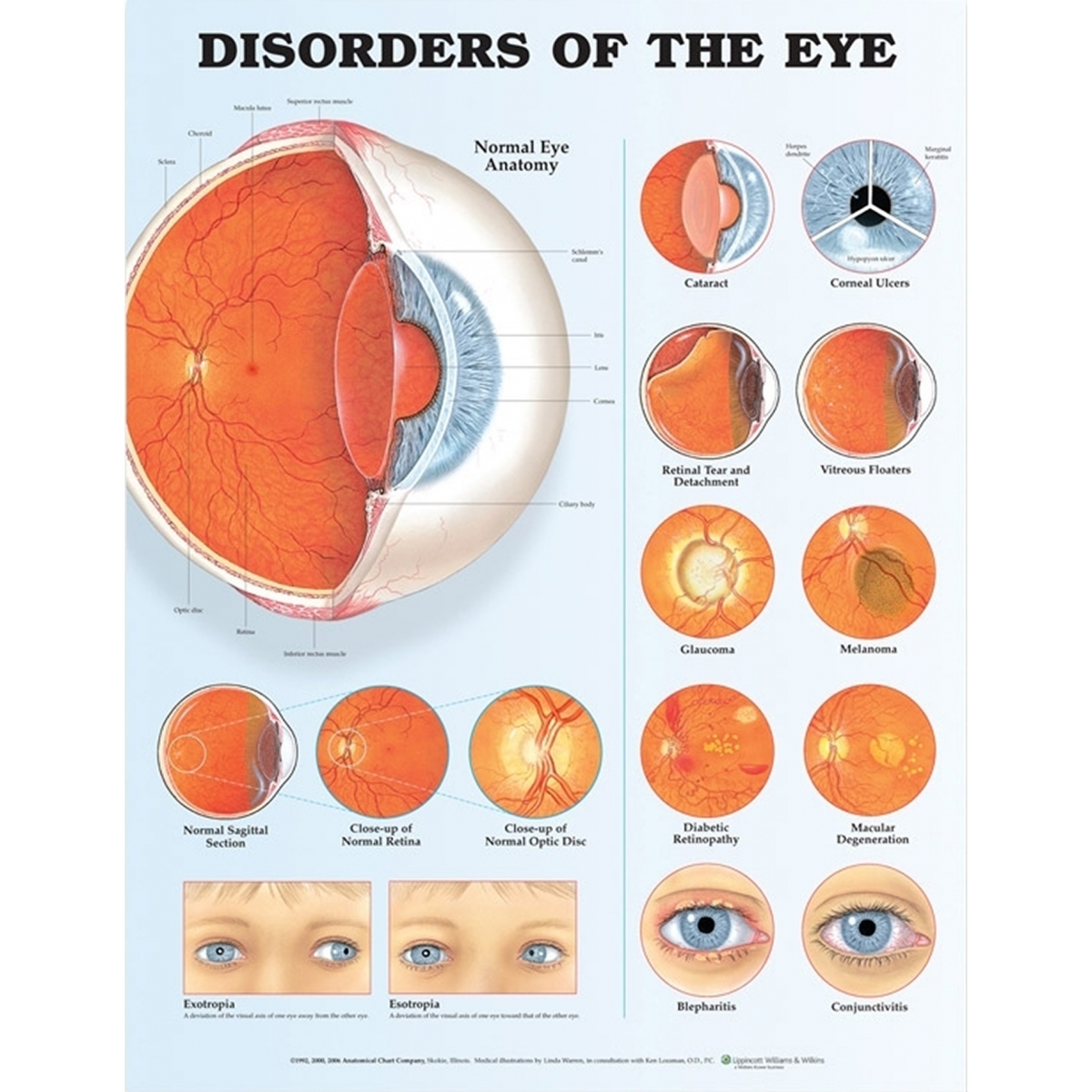
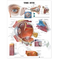
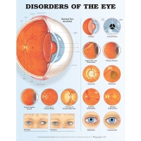
Charts (52 × 70 cm, laminated with rollers): Human Eye Anatomy and Eye Diseases & Disorders.
Learning objectives
Identify anterior/posterior segment structures and the optical pathway; relate accommodation to ciliary body–zonular mechanics.
Explain pathophysiology and clinical impact of cataract subtypes (nuclear, cortical, posterior subcapsular) and indications for IOL counselling.
Map actions and innervation of extraocular muscles (CN III/IV/VI) and correlate with strabismus testing.
Use models/charts for OSCE tasks: layer identification, pupillary reflex pathway, adnexal anatomy, and patient education.
Specifications
Material: Medical-Grade PVC.
Cleaning: mild detergent or 70% alcohol wipe; avoid solvents/heat.
Intended use: instructional aids for ophthalmology/optometry/ENT and skill-lab OSCEs.
Total Reviews (0)