


₹ 4,899 ₹ 21,199

₹ 13,899 ₹ 34,179

₹ 5,799 ₹ 7,756

₹ 5,299 ₹ 11,523
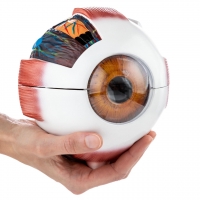
 With 7 Dissectible Parts - Myaskro®")
₹ 2,399 ₹ 7,262
 With Special Lamination")
₹ 1,599 ₹ 3,865
 With Special Lamination")
₹ 1,599 ₹ 3,865

₹ 9,299 ₹ 16,865

₹ 11,799 ₹ 18,945
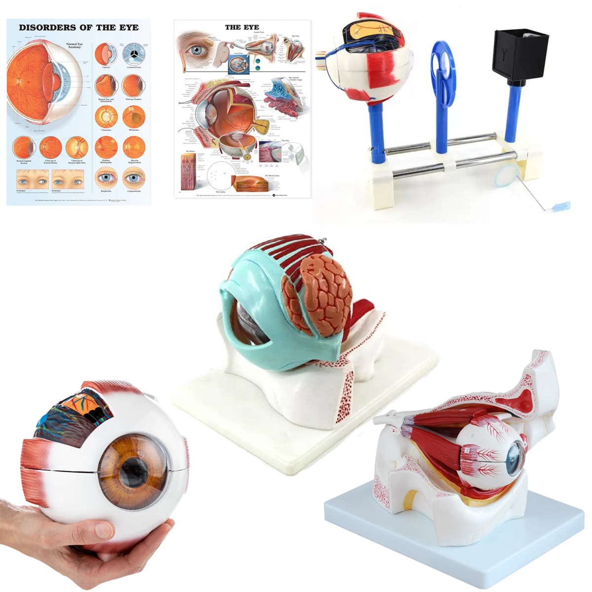
Components
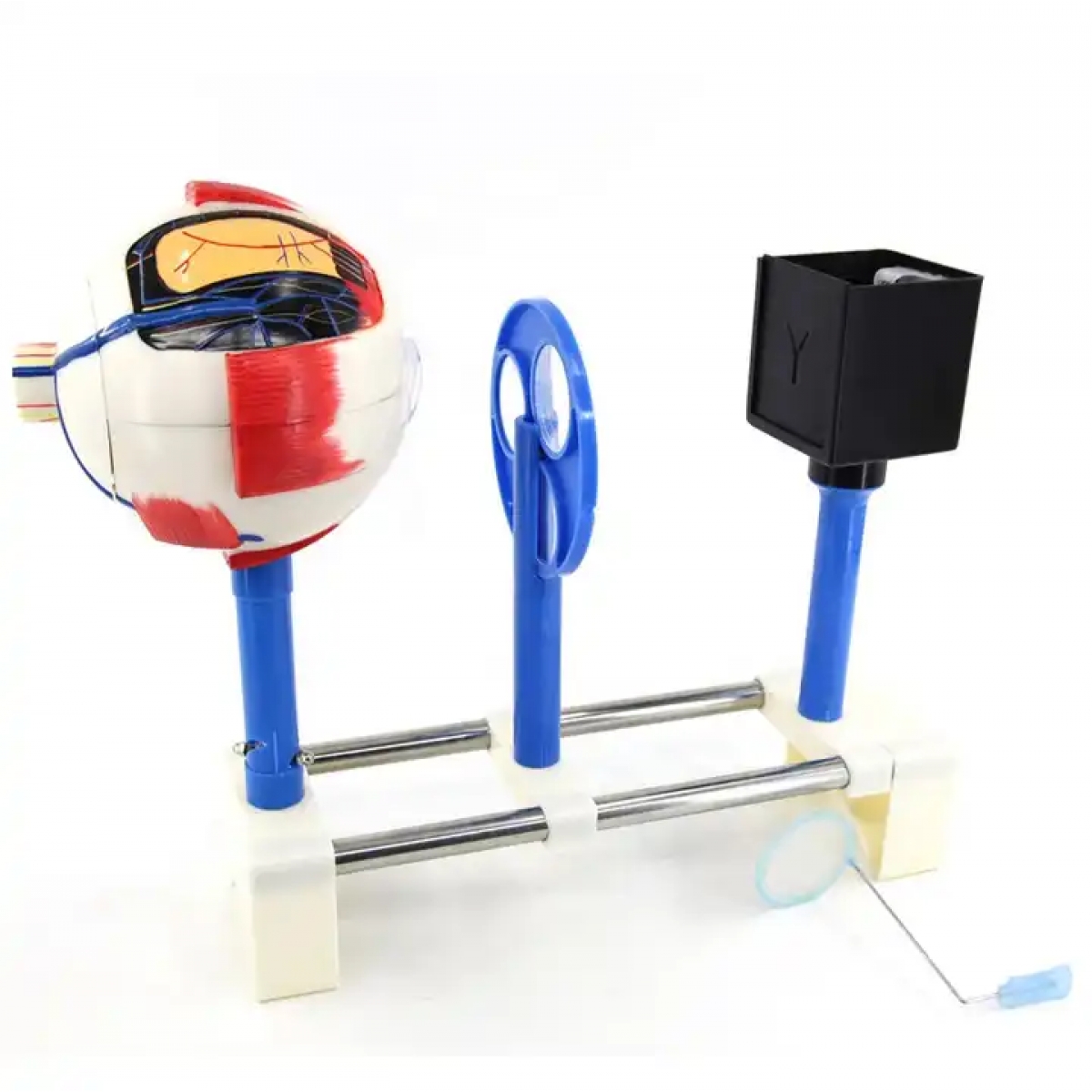
Functional human eye model: Optical bench with cornea–lens assembly, interchangeable lenses, adjustable iris diaphragm and retina screen to demonstrate emmetropia, myopia, hyperopia, astigmatism and the pinhole effect (depth of field).
Eye (6×, 6 parts): Sclera–choroid–retina shell, cornea, iris–ciliary body, crystalline lens with zonules, vitreous body, optic nerve head.
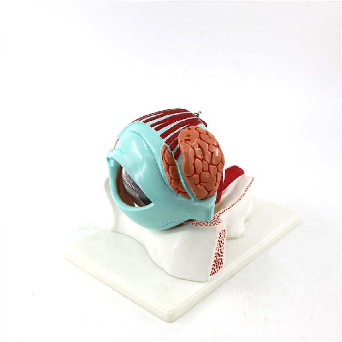
Eye with eyelid (6×, 7 parts): Above plus upper eyelid with tarsal plate and palpebral conjunctiva for lacrimal/ocular-surface teaching.
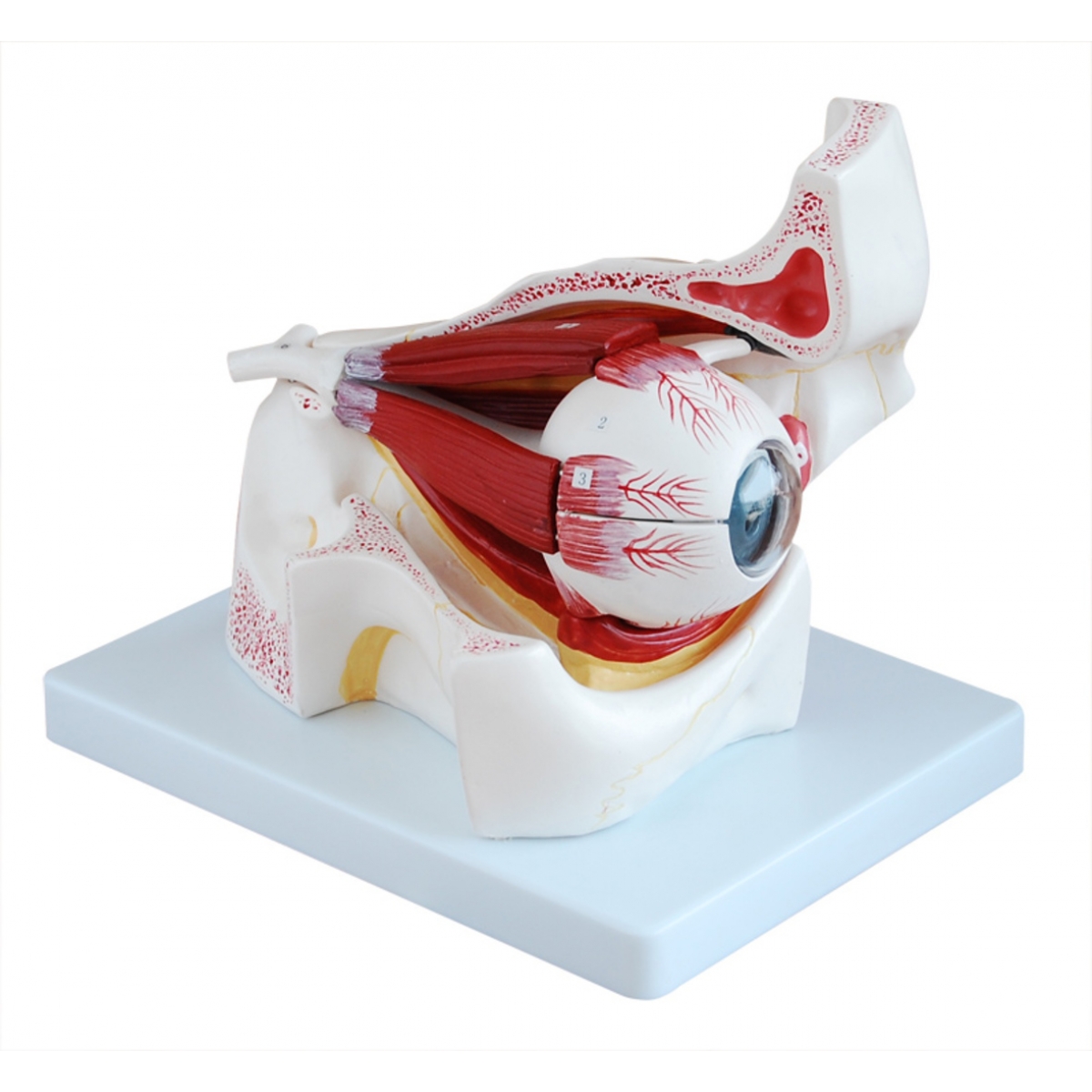
Eye with orbit (3×, 10 parts): Bony orbit with extraocular muscles (SR, IR, MR, LR, SO, IO), optic nerve, lacrimal gland region; sectional exposure of apex and fissures.
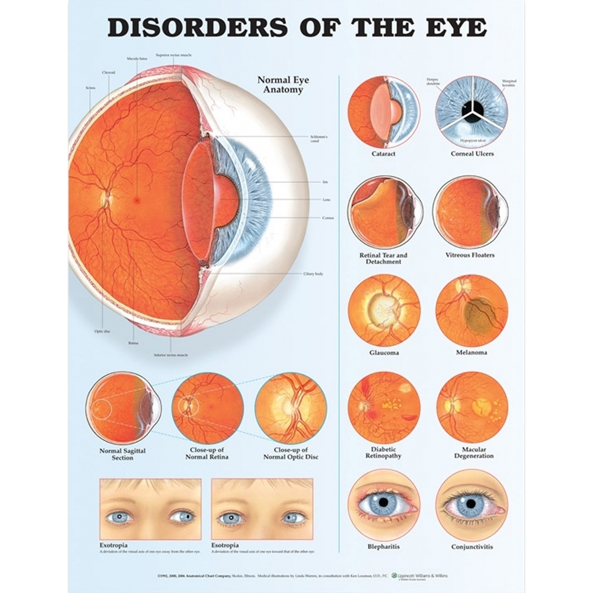
Charts (52 × 70 cm, laminated with rollers): Human Eye Anatomy and Eye Diseases & Disorders.
Learning objectives
Identify anterior and posterior segment structures: cornea, anterior chamber, iris, ciliary body, lens, vitreous, retina (macula/fovea), optic disc.
Map extraocular muscle actions and CN III/IV/VI innervation; relate to strabismus patterns.
Explain optical principles of refraction and accommodation; demonstrate correction with spherical/cylindrical lenses.
Correlate common pathology: cataract, glaucoma, diabetic retinopathy, AMD, retinal detachment, dry eye/blepharitis.
Set OSCE tasks: fundus orientation, pupillary reflex pathway discussion, ocular motility testing.
Specifications
Materials: Medical-Grade PVC.
Charts: heavy-gauge lamination; dry-wipe compatible.
Care: wipe with mild detergent or 70% alcohol; avoid solvents/heat.
Intended use: instructional aids for ophthalmology, optometry, ENT, nursing, and patient education.
Total Reviews (0)