


₹ 4,499 ₹ 6,071

₹ 5,299 ₹ 15,299
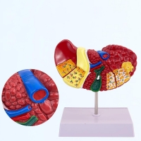
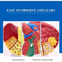
 Dissectible Into 3-Parts - Myaskro")
₹ 2,599 ₹ 7,782

₹ 6,599 ₹ 22,379

₹ 17,799 ₹ 34,155

₹ 3,299 ₹ 10,579

₹ 18,899 ₹ 26,785
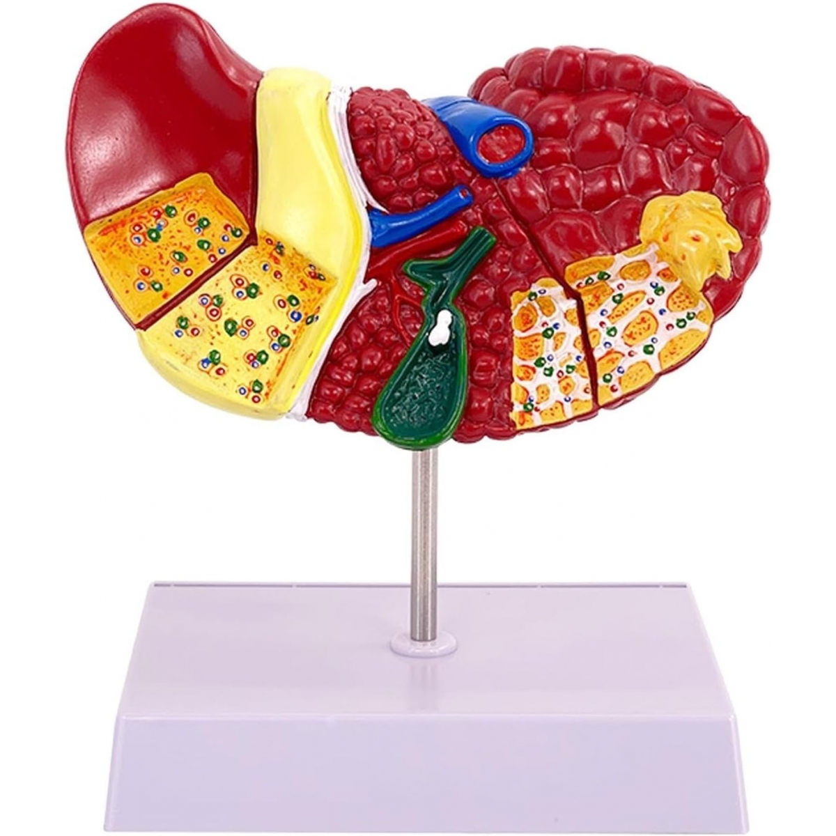
Discover a powerful new way to teach, study, and understand liver disease with the MYASKRO® Advanced Pathological Liver Model. Designed for professional-grade accuracy and clinical realism, this anatomical model provides a hands-on representation of hepatic pathology, especially cirrhosis and degenerative liver conditions. Whether you are a medical educator, a student, or a practicing physician, this model bridges the gap between textbook theory and real-world pathology.
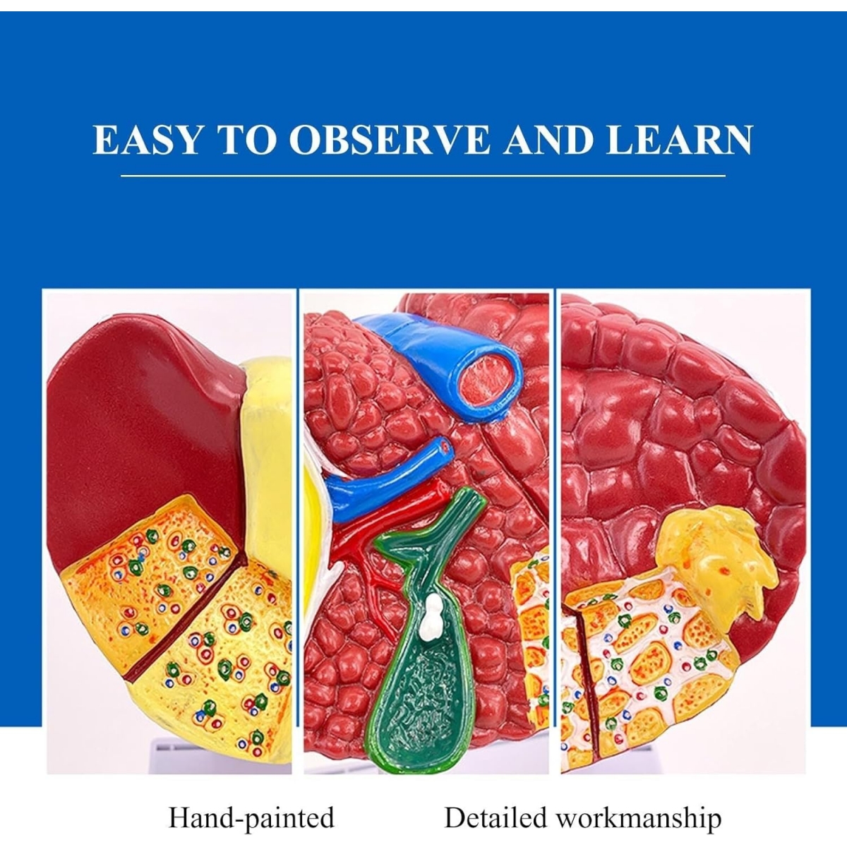
This model is not a generic liver—it is a detailed, medically accurate depiction of the pathological liver. From distorted lobules and fibrotic scarring to enlarged nodules and compressed bile ducts, the model visualizes the structural consequences of advanced liver disease in stunning three-dimensional form.
Key pathological features include:
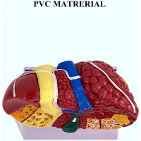
The MYASKRO® model is constructed from high-quality, medical-grade PVC plastic—the same material used in hospital training simulators. It’s lightweight, portable, and designed to withstand repeated classroom handling, clinical demonstrations, and student examinations without wear or fading.
This model was developed under the guidance of clinical pathologists and hepatologists to ensure every aspect matches real-world pathological criteria. It serves as a visual aid for lectures, patient consultations, OSCEs, and postgraduate training in gastroenterology, hepatology, and internal medicine.
Most students struggle to understand cirrhosis until they see it. This liver model makes that first encounter clear, detailed, and memorable. As students examine regenerative nodules and vascular remodeling, they connect what they’ve read with what actually occurs in clinical practice. That’s where real understanding begins.
A: Yes, this model is built to represent a life-size human liver, with exaggerated pathological features to enhance visual learning. It is perfect for demonstrating both normal anatomy and pathological deviations side by side.
A: The model includes visual representations of common and clinically significant liver conditions such as cirrhosis, fatty infiltration, fibrosis, bile duct compression, and nodular regeneration. It gives a complete view of disease progression from healthy to advanced pathological states.
A: This particular model is a non-detachable single unit, securely mounted on a stand. It is designed for visual inspection, touch-based exploration, and presentation—not for internal dissection.
A: It is ideal for medical and nursing students, teaching professionals, clinical instructors, practicing physicians, and healthcare trainers. Additionally, it is used in hospital patient rooms for clear and effective disease explanation during consultations.
A: Yes, simply use a soft, damp cloth to wipe the surface. The PVC material resists staining, discoloration, and deterioration—so it’s built to look new even after years of use.
What sets this model apart is its dual benefit—it is as effective in a teaching hall as it is in a doctor’s office. Professors appreciate its detail; students value its clarity; physicians trust it as a visual aid during patient consultations.
“This model simplifies complex pathology. It helps students understand cirrhosis in minutes, not hours. I use it in every class I teach.”
– Dr. R. Malhotra, Professor of Hepatology
“During patient consultations, it’s invaluable. You can explain liver disease progression with clarity and impact—no more confusion or miscommunication.”
– Dr. Sameer Kulkarni, Internal Medicine Consultant
MYASKRO® is a name trusted by hundreds of medical institutions, universities, and healthcare educators across the country. We don’t just make models—we design educational tools with a single goal: to improve understanding through clarity, quality, and clinical realism.
Total Reviews (0)