

Anatomic Details
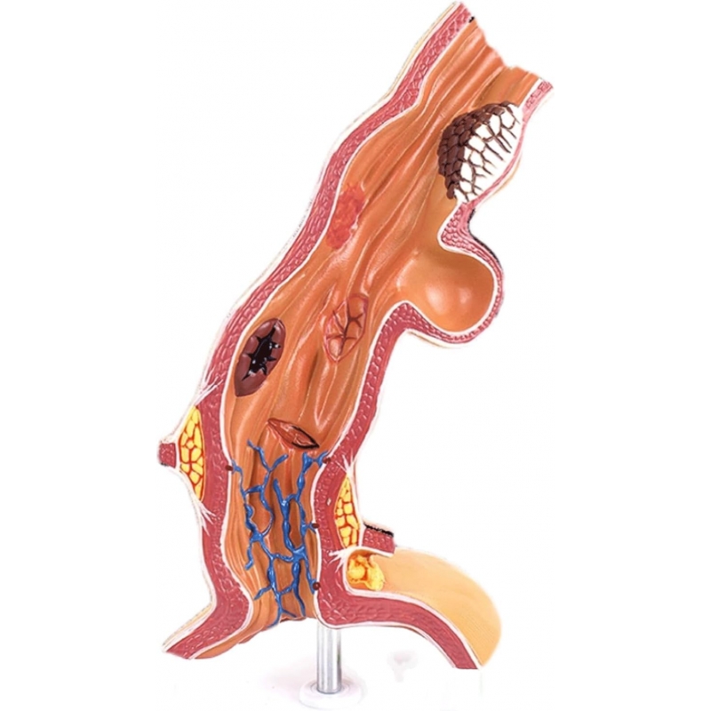
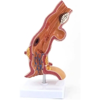
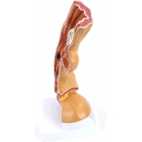
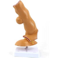
Enlarged longitudinal section of the esophagus demonstrating mucosa (non-keratinized squamous epithelium), submucosa with venous plexus, muscularis propria (inner circular/outer longitudinal), and outer connective tissue.
Distal squamocolumnar junction (Z-line) and lower esophageal sphincter (LES) region for reflux discussion.
Representative lesions: erosions/ulceration, esophageal varices, diverticulum, and malignant mass infiltration.
Learning objectives
Differentiate esophageal wall layers and correlate with endoscopic appearances.
Explain pathophysiology of GERD/erosive esophagitis, variceal formation, diverticular disease, and esophageal carcinoma.
Use in OSCEs for lesion identification and counselling on alarm symptoms/dysphagia.
Specifications & use
Medical Grade PVC.
Wipe with mild detergent or 70% alcohol; avoid solvents/heat.
For UG/PG anatomy, gastroenterology, GI surgery, nursing, and patient education.
Total Reviews (0)