


₹ 2,199 ₹ 10,281

₹ 6,800 ₹ 15,157

₹ 1,699 ₹ 7,251

₹ 3,399 ₹ 7,613

₹ 8,699 ₹ 19,420
 , 26cm Tall - MYASKRO")
₹ 2,999 ₹ 9,611
See the Science. Teach the Detail.
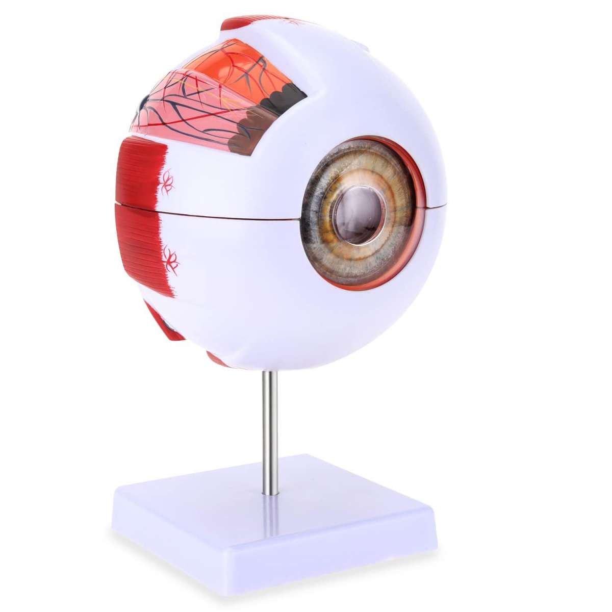
The MYASKRO® Human Eye Model is an anatomically accurate, 6-times enlarged replica designed to show every intricate structure of the human eye with crystal clarity. Built with 7 dissectible components, this educational tool is ideal for science teachers, ophthalmology instructors, medical students, and healthcare educators who want a hands-on, visual way to demonstrate eye anatomy and function.
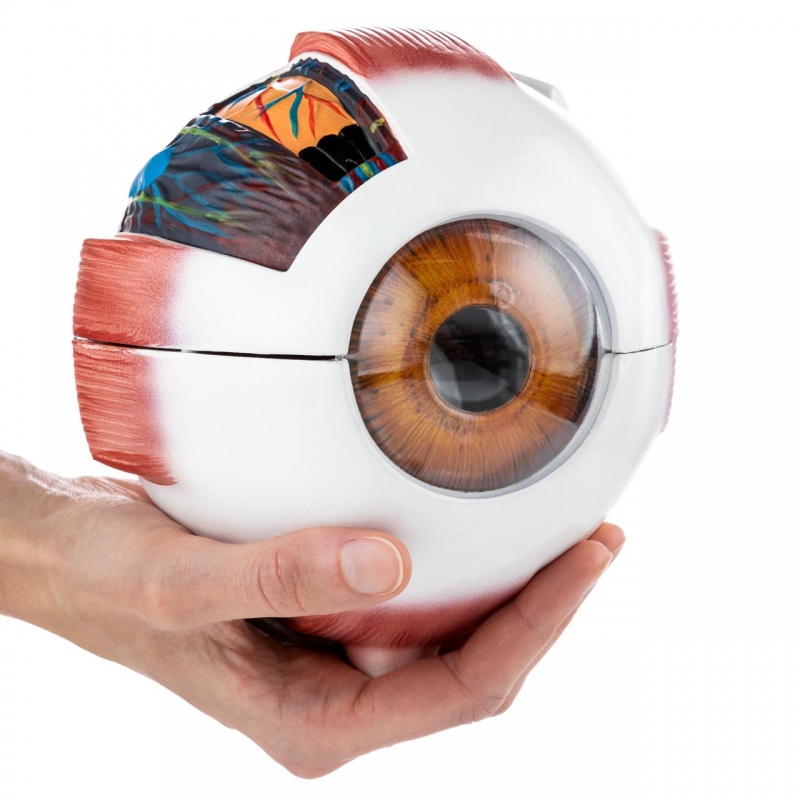
The human eye is incredibly small—and incredibly complex. With over a dozen key internal structures packed into a sphere roughly 2.5 cm in diameter, it's a challenge to teach its function and structure without magnification. This model solves that problem by enlarging the entire eye 6 times, allowing every nerve, muscle, and chamber to be seen, named, and understood.
Educators use this model to walk students through the visual process, from the moment light enters the eye to when a signal reaches the visual cortex. The detachable design allows instructors to pass parts around, quiz students on functions, and engage multiple senses for deeper learning. The enlarged size is ideal for lecture halls, demonstration tables, and hands-on practical exams.
The human eye is an organ of extraordinary complexity, but MYASKRO® makes it teachable. The layered build, accurate coloring, and tactile learning style help students retain concepts far beyond the classroom.
This model is more than a prop—it’s an interactive learning experience. Use it to simplify exams, reinforce lectures, and empower your students with knowledge they can literally hold in their hands.
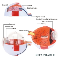
Visual and tactile tools improve retention dramatically. In fact, educational research shows that learners retain up to 60% more information when lessons are supported with 3D models. That’s especially true in anatomy and physiology, where abstract terms like “retina” or “ciliary body” become real when they can be seen, touched, and reassembled by hand.
For instructors, this model brings lectures to life. For students, it transforms memorization into mastery.
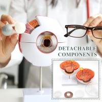
The MYASKRO® eye model disassembles cleanly into seven pieces. Each part is easy to remove and reposition without tools or force. The internal structures align snugly when reassembled, making the model ideal for demonstration, discussion, and repetition.
Because it’s mounted on a stable base, the model can be rotated and positioned at any angle—ideal for desks, podiums, or lab benches. Pass it around the classroom, display it in clinics, or use it one-on-one to explain patient conditions.
Constructed from high-quality PVC and hand-painted with medical-grade dyes, the MYASKRO® eye model is built for real-world educational environments. It’s:
No. It is deliberately enlarged to 6 times the size of a real human eye for clearer visual instruction.
Yes. The model is designed for repeated disassembly and reassembly. All 7 parts fit securely and align anatomically when assembled.
This model is ideal for middle school through university-level students. It’s safe for classroom or clinic use under supervision.
Absolutely. It’s perfect for ophthalmologists, optometrists, and general physicians who need to explain eye surgeries, vision disorders, or treatment plans in a visual, relatable way.
“This model made our optics class so much easier to teach. Students immediately grasp how light enters and bends.”
– Prof. Anurag M, Anatomy Instructor“Perfect for exam prep. Took it apart and reassembled it 20 times before my OSCE. I’ll never forget the eye’s structure now.”
– Priya K., Med Student“Explaining glaucoma used to take 10 minutes. With this model, I show it in 30 seconds. Patients get it.”
– Dr. Rehman, Ophthalmologist
Master the structure of sight. Whether you're a teacher, student, or healthcare professional, the MYASKRO® Enlarged Human Eye Model gives you the clarity to teach, learn, and communicate with impact.
Total Reviews (0)