


₹ 4,199 ₹ 15,063

₹ 1,999 ₹ 5,427

₹ 3,899 ₹ 9,316

₹ 1,499 ₹ 4,443

₹ 4,799 ₹ 7,315

₹ 5,599 ₹ 9,622
 - MYASKRO")
₹ 4,699 ₹ 8,431

₹ 8,499 ₹ 25,919

₹ 2,899 ₹ 4,537

₹ 3,599 ₹ 6,712
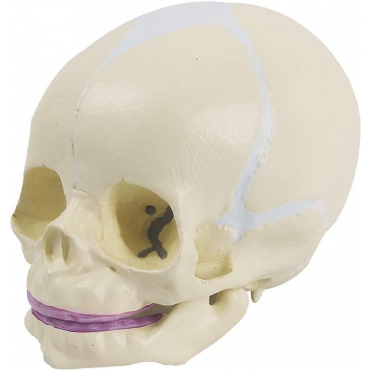
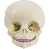
The human skull is a marvel of structural design—and nowhere is it more delicate, adaptable, and instructive than in its earliest stage of life. The MYASKRO Fetal Skull Model captures this moment in stunning anatomical precision, offering students, clinicians, and educators an invaluable look at the prenatal and neonatal cranial structure.
This life-sized model accurately depicts the fontanelles, cranial sutures, and flexible joint lines that define a fetal skull’s unique morphology. Whether you're teaching embryology, neonatology, or midwifery, this model reveals the intricate balance between rigidity and pliability—essential to both brain protection and natural childbirth.
Constructed from durable, medical-grade PVC, the model feels lifelike yet resilient, suitable for frequent handling in classrooms, clinical simulations, and one-on-one patient education. The delicate cranial bones are color-enhanced to help distinguish between sutures, growth plates, and developing bone margins at a glance.
Key Features:
It’s especially effective in teaching:
As one anatomy instructor commented, “This model brings clarity to one of the most misunderstood areas of pediatric anatomy. You don’t fully appreciate how the skull accommodates birth and brain growth until you’ve held this in your hand.”
Compact, educational, and visually striking, the MYASKRO Fetal Skull Model delivers not just structure—but story. It tells how our most complex organ—the brain—is protected, supported, and nurtured from the very beginning.
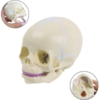
The MYASKRO Fetal Skull Model goes beyond bone—it’s a map of how life adapts to grow, survive, and pass through the most physically demanding event of early existence: childbirth. With its open fontanelles and visible cranial sutures, this model teaches more than structure; it explains function, flexibility, and clinical relevance in a way few models can.
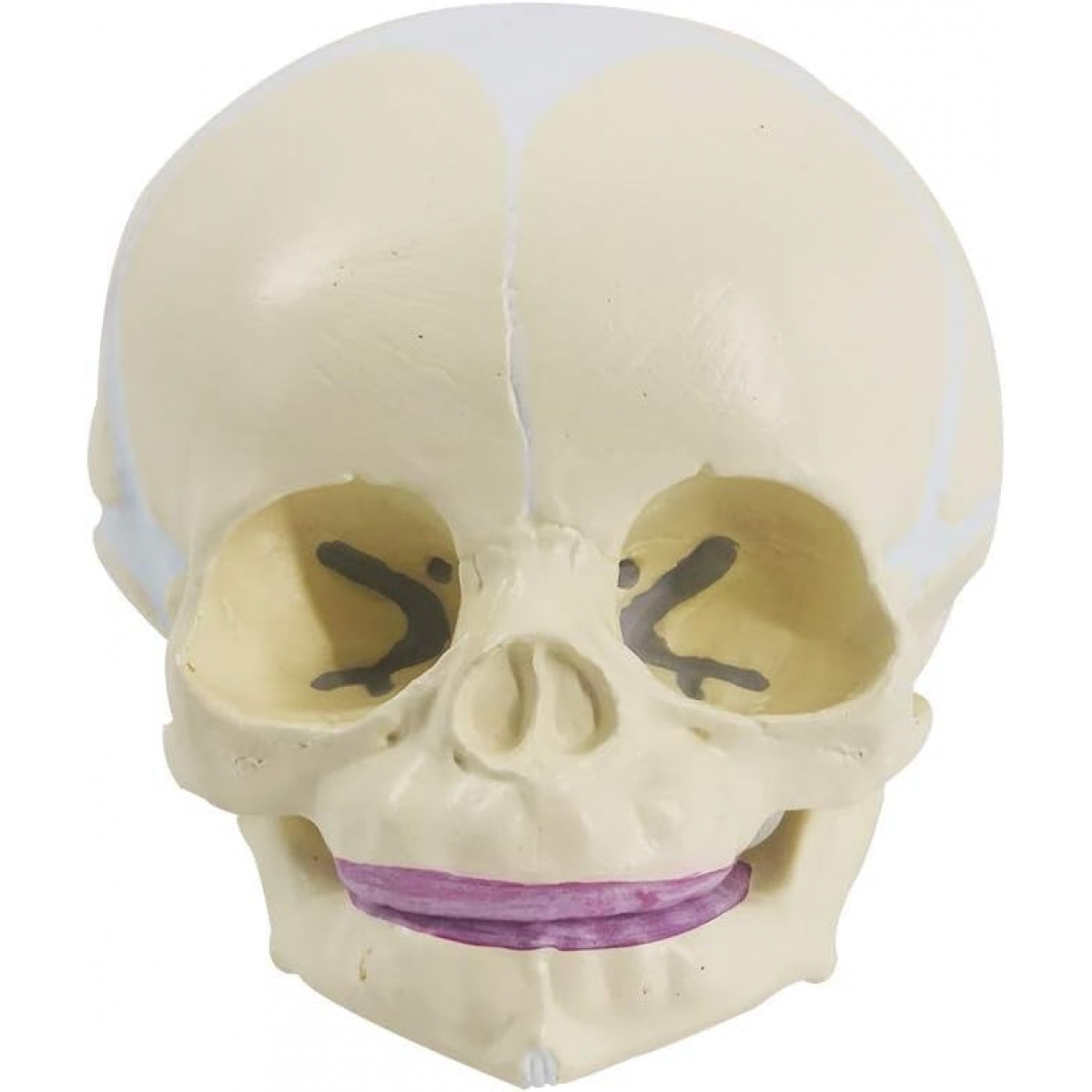
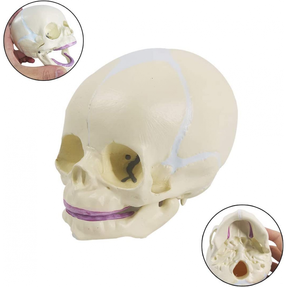
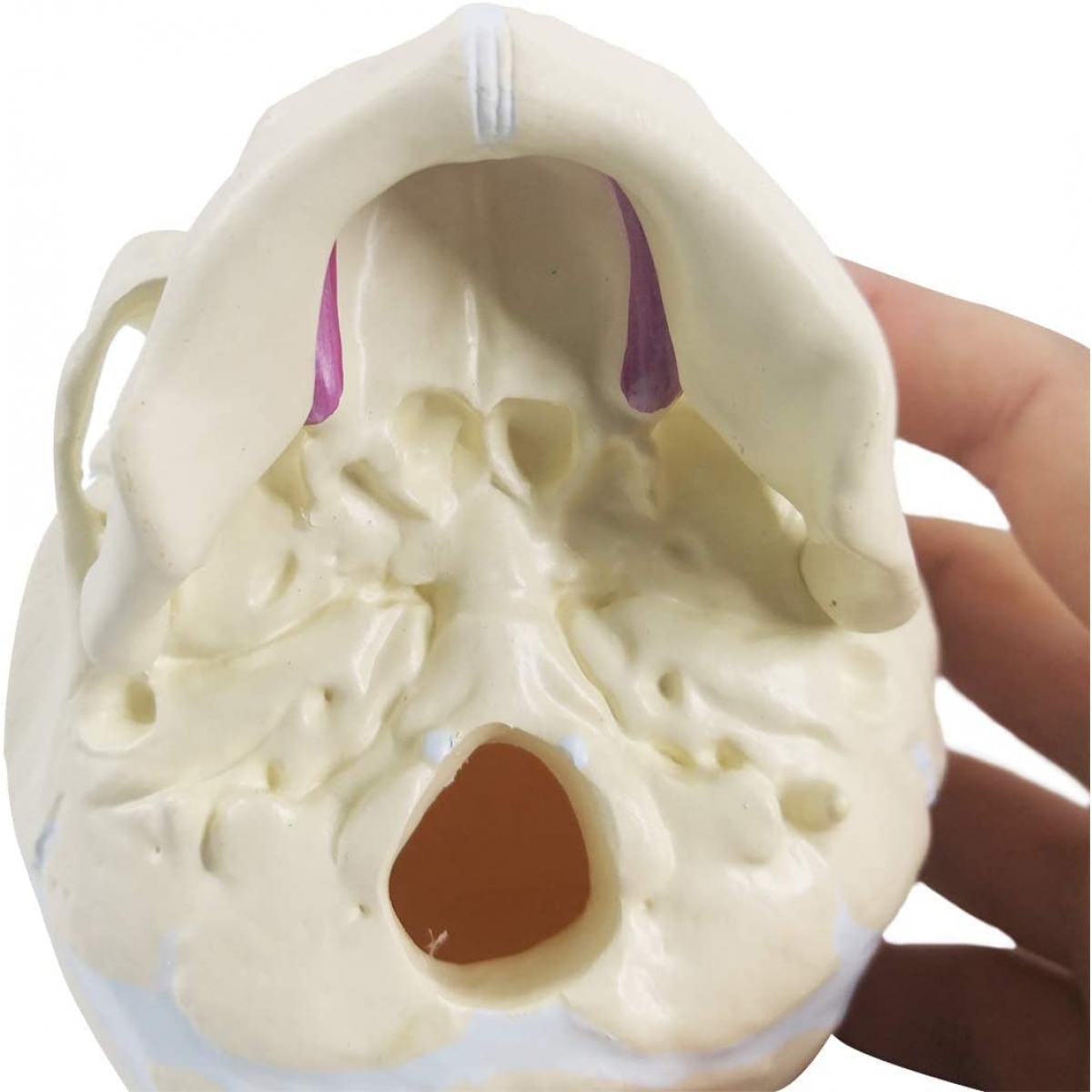
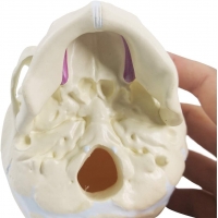
At birth, the human skull is unlike any other time in life. The bones aren’t yet fused, and the gaps—known as fontanelles—are key to both delivery and brain development. This model makes it easy to identify the anterior, posterior, sphenoidal, and mastoid fontanelles, each delicately molded and colored to enhance visibility without compromising realism.
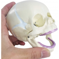
Students can trace the sagittal suture running down the midline, meet the coronal suture separating frontal and parietal bones, and examine the lambdoid and squamosal sutures along the posterior and lateral skull. These anatomical details aren’t just cosmetic—they’re critical for understanding how the skull plates overlap (a process called molding) during vaginal delivery.
Midwifery instructors often use this model during labor and delivery modules, showing how palpation of fontanelles during internal exams helps assess fetal position. Pediatricians and child health educators benefit from being able to illustrate normal cranial development and address concerns like plagiocephaly or delayed suture closure. Dental educators will also appreciate the inclusion of emerging deciduous teeth in the upper and lower gum pads—an excellent touch for teaching oral development in infants.
The model’s size and portability make it ideal for table-top demonstration or bedside explanation. Weighing under 250 grams, it fits easily into teaching kits, anatomy labs, or clinic displays. And unlike some fetal models that lose anatomical fidelity for size, this one maintains clinical accuracy while being compact enough for focused study.
Made of premium PVC, the model is easy to clean and durable enough to survive years of classroom use. Students can safely pass it around, trace the sutures, and quiz themselves on fontanelle landmarks—building both familiarity and confidence in early cranial anatomy.
In the words of one medical student, “It helped me visualize how soft and mobile the fetal skull really is. Suddenly, the concept of molding during birth made perfect sense.”
For anyone teaching or learning about childbirth, pediatric care, or early skull development, this model offers a clarity that’s hard to replicate with diagrams alone.
The MYASKRO Fetal Skull Model may be compact, but the depth of understanding it offers is anything but small. This model bridges the gap between textbook anatomy and real-world clinical insight, allowing learners to grasp how the fetal skull supports both neurodevelopment and safe passage through the birth canal. It’s a teaching tool that clarifies one of the most dynamic stages of human skeletal evolution—where growth, adaptation, and protection intersect.
For students in obstetrics, midwifery, pediatrics, or craniofacial medicine, the model functions as a visual and tactile guide through the subtleties of fetal anatomy. The soft, unfused skull with distinct suture lines and fontanelles reflects nature’s perfect compromise between structure and flexibility. The bones aren’t rigid—they're engineered for movement, shaping, and rapid brain expansion during infancy. This makes understanding the fetal skull not just helpful, but foundational to every healthcare professional who will care for newborns.
Educators find particular value in this model for palpation training and anatomical orientation. Instructors can simulate head positioning during labor, teach the difference between anterior and posterior fontanelles, or demonstrate how overlapping sutures aid delivery. When paired with pelvic models, it enhances simulation scenarios and makes labor mechanics easier to conceptualize.
Parents, too, benefit when clinicians use this model for patient education. Whether discussing normal newborn head shape, craniosynostosis, or simply explaining why babies have “soft spots,” a physical model offers clarity and reassurance that printed brochures rarely provide. It's a gentle, approachable way to engage in conversations that matter deeply during those first days of life.
The model’s durable PVC material ensures it holds up to frequent use, while its life-sized proportions maintain realism without sacrificing portability. And thanks to the clear contrast of painted sutures and gum pads, learners at any level—visual, tactile, or kinesthetic—will find it easier to engage, retain, and apply the knowledge it represents.
As one clinical educator noted, “This model makes teaching neonatal skull anatomy far more interactive and effective. It invites curiosity, fosters discussion, and strengthens clinical confidence.”
In medical education, the best tools don’t just inform—they illuminate. The MYASKRO Fetal Skull Model does exactly that, helping learners and professionals alike understand not just the bones of the skull, but the brilliant biological design that allows both birth and brain growth to happen in harmony.
Total Reviews (0)