 With Femur Heads - MYASKRO")
₹ 1,399 ₹ 6,241
|MYASKRO")
₹ 9,299 ₹ 14,545

₹ 3,999 ₹ 7,315
")
₹ 6,499 ₹ 9,982
 - MYASKRO")
₹ 7,499 ₹ 23,453
")
₹ 7,799 ₹ 12,845

₹ 11,799 ₹ 18,945
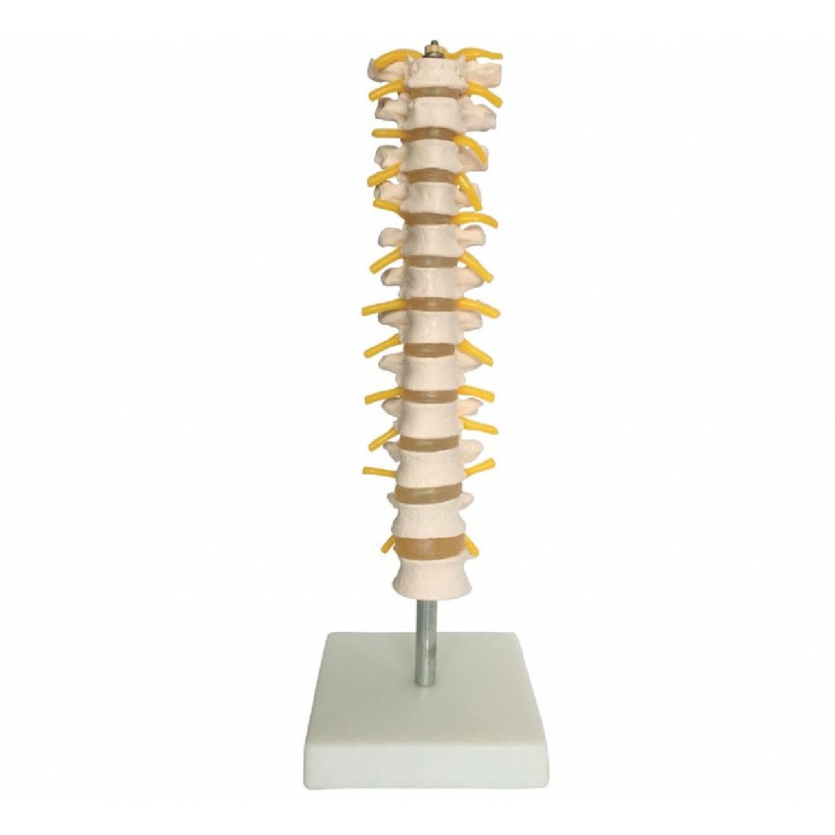
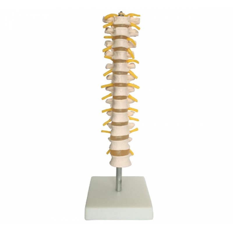
The thoracic spine plays a pivotal role in structural stability, rib articulation, and spinal nerve distribution. Understanding its form is essential for anyone working in orthopedics, physiotherapy, chiropractic care, or anatomy education. The MYASKRO Thoracic Spinal Column Model delivers an accurate, full-scale representation of this critical region—designed to bring clarity to complex concepts.
This life-size model features T1 to T12 vertebrae, complete with intervertebral discs, spinous and transverse processes, and yellow nerve root projections emerging between each segment. Mounted on a solid base for stability, it provides a tactile and visual experience ideal for students, practitioners, and patient education.
Whether you're explaining spinal alignment to a patient or preparing an MBBS batch for practical exams, this model gives you the ability to demonstrate anatomy with hands-on precision. Each vertebra is cast from high-grade PVC for clinical accuracy, long-term durability, and ease of handling.
Used widely in physiotherapy colleges, spine clinics, orthopedic OPDs, chiropractic centers, and classroom settings, this model helps connect textbook knowledge to real-world understanding. It’s especially valuable for discussing conditions like kyphosis, thoracic disc compression, postural deviations, or scoliosis.
Relevant search queries like “thoracic spine anatomy model,” “T1–T12 vertebrae model,” “PVC spine column model India,” and “educational thoracic vertebrae replica” lead directly to this clinical-quality product.
“This model helped our BPT students at Manipal University grasp spinal nerve orientation and thoracic mobility limitations within minutes,” says Dr. Prerna Chhabra, Lecturer in Anatomy.
If you teach, diagnose, or rehabilitate anything related to thoracic function, this model doesn’t just assist—it elevates your ability to communicate spinal anatomy with unmistakable clarity.
The MYASKRO Thoracic Spinal Column Model offers a deep dive into the anatomy and biomechanics of the mid-back. Representing vertebrae T1 to T12, this model is crafted to deliver exceptional anatomical precision and educational value.
Each vertebra is molded to highlight:
This model is ideal for demonstrating how thoracic structure contributes to:
Educators in MBBS, BPT, and allied health programs use this tool to reinforce practical knowledge before students ever touch a cadaver or real patient. Likewise, clinicians rely on it to explain spinal misalignments, nerve impingement, and mid-back conditions in a way patients can visualize and trust.
Medical trainers often ask: “Can it be used for comparative spine studies?” Yes. This model pairs excellently with cervical or lumbar sections to illustrate differences in vertebral structure and curvature along the spine.
Made from non-toxic, high-density PVC, this model is both lightweight and built to withstand regular handling in academic or clinical settings. The base is solid, ensuring stability on a consultation desk or lab bench. The height and width proportions match adult thoracic anatomy, providing a truly life-size reference.
Search-aligned keywords include “T-spine vertebral column model,” “thoracic nerve exit model,” “PVC thoracic vertebrae teaching tool,” and “spinal column model India.”
“At our chiropractic campus in Pune, this model is used to teach everything from nerve root compression to vertebral body palpation,” shares Prof. Ujjwal Mehta, Academic Coordinator.
Whether you’re explaining anatomy or pathology, this model turns abstract spinal concepts into something concrete, visual, and unforgettable.
The thoracic region is often overlooked in favor of the cervical or lumbar spine, yet it's essential for understanding posture, rib function, and spinal nerve distribution. The MYASKRO Thoracic Spinal Column Model gives this middle segment of the vertebral column the attention—and accuracy—it deserves.
Whether you’re preparing for practical exams, teaching a classroom full of MBBS aspirants, or guiding patients through thoracic spine-related conditions, this model gives you the confidence and clarity to explain with precision. Every spinous notch, transverse process, and nerve root tells a story, and this model helps you narrate it effectively.
Its compact size makes it ideal for portable use in clinical settings or educational demonstrations, yet the level of anatomical detail rivals full-body spine models. With the nerve roots painted in high-visibility yellow, it becomes even easier to highlight the connection between thoracic vertebrae and intercostal nerve pathways—a critical detail in physiotherapy and neurology education.
For institutions looking to elevate their teaching standards, this model offers a cost-effective, space-saving, and accurate solution. It’s already in use across physiotherapy departments, spine care clinics, radiology labs, and orthopedic teaching hospitals throughout India.
This model also meets strong online visibility benchmarks for terms like “T1-T12 vertebrae anatomical model,” “spinal nerve demonstration spine,” “thoracic vertebral column PVC model,” and “mid-back spine model for education.” It meets the expectations these searches imply—clinical realism, functional design, and academic utility.
“Explaining thoracic pain patterns to patients used to take five minutes—now it takes thirty seconds with this model,” says Dr. Ananya Joshi, Spine Specialist, Apollo Clinic, Ahmedabad.
If your goal is to make spinal anatomy simple, visible, and teachable, then this model is more than just a prop—it’s a professional tool. Quiet in form, powerful in impact, and trusted by India’s medical educators and practitioners alike.
Total Reviews (0)