

₹ 3,599 ₹ 6,712

₹ 21,299 ₹ 45,979
 - Myaskro")
₹ 5,699 ₹ 15,181
 - Myaskro")
₹ 5,699 ₹ 15,299

₹ 7,499 ₹ 22,379

₹ 3,899 ₹ 9,316

₹ 3,999 ₹ 6,700

₹ 1,799 ₹ 4,301

₹ 3,999 ₹ 6,094

₹ 1,499 ₹ 4,891
 With Femur Heads & Pelvis")
₹ 5,499 ₹ 11,617
 - Myaskro")
₹ 148,699 ₹ 293,767
")
₹ 7,799 ₹ 12,845
")
₹ 6,999 ₹ 14,845
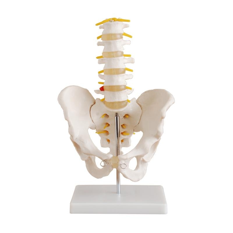
Understanding human biomechanics starts with the spine and pelvis—the architectural foundation of movement, balance, and posture. The MYASKRO Pelvis with 5 Lumbar Vertebrae model offers an unparalleled opportunity to study this critical structure in precise, life-size detail. It’s a favorite among physiotherapists, orthopedic educators, medical students, chiropractors, and osteopaths for one reason: it turns complex spinal anatomy into something tactile, teachable, and clear.
This model features five lumbar vertebrae, each with visible intervertebral discs, spinal nerves, and a detailed sacrum—mounted on a stable base. The pelvic bones are carefully molded to represent anatomical accuracy, from the iliac crest to the pubic symphysis. Together, this setup provides a visual and physical reference point that reinforces theory through interaction.
In a classroom or clinic, it bridges the gap between diagrams and diagnostics. Whether demonstrating disc herniation, spinal loading, nerve impingement, or hip alignment issues, this model supports clinical explanation and academic instruction alike. It becomes a conversation starter for patients, a test prep essential for students, and a demonstrative aid for physical therapy routines.
Wondering if the size matches reality? It does. This is a true-to-scale anatomical replica, giving users the ability to understand spatial relationships between vertebrae, sacrum, and pelvic landmarks with real-world proportions. From the transverse process of L1 to the sciatic notch, each structure is crafted to mirror its real-life counterpart.
Relevant search queries like “pelvis and spine model,” “life-size lumbar vertebrae model,” “anatomical spine model with nerves,” and “orthopedic teaching tools” consistently lead users here. And for good reason—the model satisfies professional expectations without the inflated price tag.
“I’ve taught orthopedic biomechanics for 14 years, and this is hands-down the most effective pelvis-spine model I’ve used. Accurate, durable, and student-friendly,” says Dr. Jordan R., Orthopedic Educator, Manchester.
If your goal is to improve understanding, reduce guesswork, and elevate your teaching or consultation sessions, this model belongs on your desk. It’s more than a static display—it’s a tool that brings anatomy to life with every vertebra and every nerve fiber visible in 3D clarity.
The MYASKRO Pelvis with 5pcs Lumbar Vertebrae delivers realistic, hands-on access to the structural core of human movement. Every curve, notch, and disc has been reproduced to facilitate visual understanding and tactile learning. With this model, you don’t just study the lumbar spine—you feel how it functions, segment by segment.
The vertebrae are fully molded to show spinous processes, vertebral bodies, transverse processes, and intervertebral discs. Yellow spinal nerves emerge naturally between vertebrae, allowing users to demonstrate impingement scenarios and nerve root compression with ease—perfect for neurology and orthopedics education.
Chiropractors often ask: “Can this show misalignment or disc degeneration?” While static in nature, the design allows for accurate explanation of vertebral positioning and disc space changes. With the ability to highlight common issues like lumbar lordosis, spinal stenosis, and sacroiliac dysfunction, this model is a reliable reference point in consultations and education alike.
Physiotherapy programs use it to show lumbar-pelvic stability relationships. Medical schools use it for neuroanatomy labs. Even fitness professionals leverage it to educate clients on lifting mechanics and back care. Wherever posture, pain, or pelvic balance are discussed, this model becomes the demonstrative standard.
Constructed from a medical-grade PVC blend, the model resists damage and discoloration—even with repeated classroom handling. Its mounted base provides display stability, while the modular vertebrae and pelvis make it easy to transport for off-site workshops or mobile instruction.
Instructors benefit from the clarity. Students benefit from the scale. And patients benefit from a clearer understanding of their spinal health. It’s no surprise that queries like “lumbar spine teaching model,” “sciatic nerve model,” “pelvic structure for PT education,” and “chiropractic spine replica” consistently lead to this trusted teaching tool.
“Every detail’s there—from the nerve roots to the intervertebral spacing. It’s helped my students visualize lumbar dysfunction in a way 2D diagrams never could,” says Melissa V., PT Instructor, Toronto.
Whether you're mapping nerve pathways or explaining chronic back pain, this model supports clear, confident communication. It’s the kind of tool that makes complex anatomy feel a little simpler—and far more approachable.
True anatomical understanding comes from repetition, demonstration, and engagement—and the MYASKRO Pelvis with 5 Lumbar Vertebrae offers all three in one dependable tool. It’s a no-nonsense model with just the right amount of complexity to satisfy professionals, without overwhelming beginners.
For anatomy instructors, it’s an intuitive way to explain the lumbosacral junction, nerve pathways, and postural alignment. For clinicians, it becomes a communication bridge when explaining back pain, spinal conditions, or surgical options to patients. And for students, it serves as a tangible memory aid—anchoring theoretical knowledge in something physical and real.
Wondering if it's limited to orthopedic use? Not at all. This model fits into disciplines ranging from physical therapy, sports science, osteopathy, massage therapy, midwifery, chiropractic, and general medicine. If your work involves the lower spine, pelvis, or nerve roots, this model becomes a daily asset.
And its portability makes it versatile. You can take it from the classroom to the consultation room. From the anatomy lab to the training hall. From a university lecture to a workshop demonstration. Its sturdy base and detachable construction make setup effortless and transport stress-free.
It also earns praise for its visual clarity. The contrasting nerve color, defined vertebral architecture, and anatomical landmarks allow observers to absorb information quickly—especially helpful in fast-paced teaching environments or patient consultations where time is limited.
Semantic alignment? Absolutely. Users searching terms like “spine and pelvis anatomy model,” “lumbar disc and nerve trainer,” “L1–L5 vertebrae replica,” or “pelvic model with spine for teaching” will find this tool ranking where it matters—because it consistently delivers on those expectations.
“We keep one in every lab. It’s the fastest way to explain posture, disc mechanics, or nerve impingement. Everyone—from students to patients—understands better when they see it,” says Dr. Nina P., Biomechanics Lecturer, Berlin.
In the end, it’s not about having a model—it’s about having the right model. One that holds up under use, clarifies the complex, and brings anatomy out of the abstract. That’s exactly what the MYASKRO spine and pelvis model does—with quiet reliability and expert precision.
Total Reviews (0)