

₹ 5,799 ₹ 18,761

₹ 12,499 ₹ 24,965
|MYASKRO")
₹ 9,299 ₹ 14,545
 With Femur Heads - MYASKRO")
₹ 1,399 ₹ 6,241

₹ 1,299 ₹ 5,717

₹ 1,899 ₹ 2,241
 Set Anatomical Model - MYASKRO")
₹ 1,899 ₹ 3,357
 - MYASKRO")
₹ 7,499 ₹ 23,453
 With Femur Heads & Pelvis")
₹ 5,499 ₹ 11,617
")
₹ 7,799 ₹ 12,845
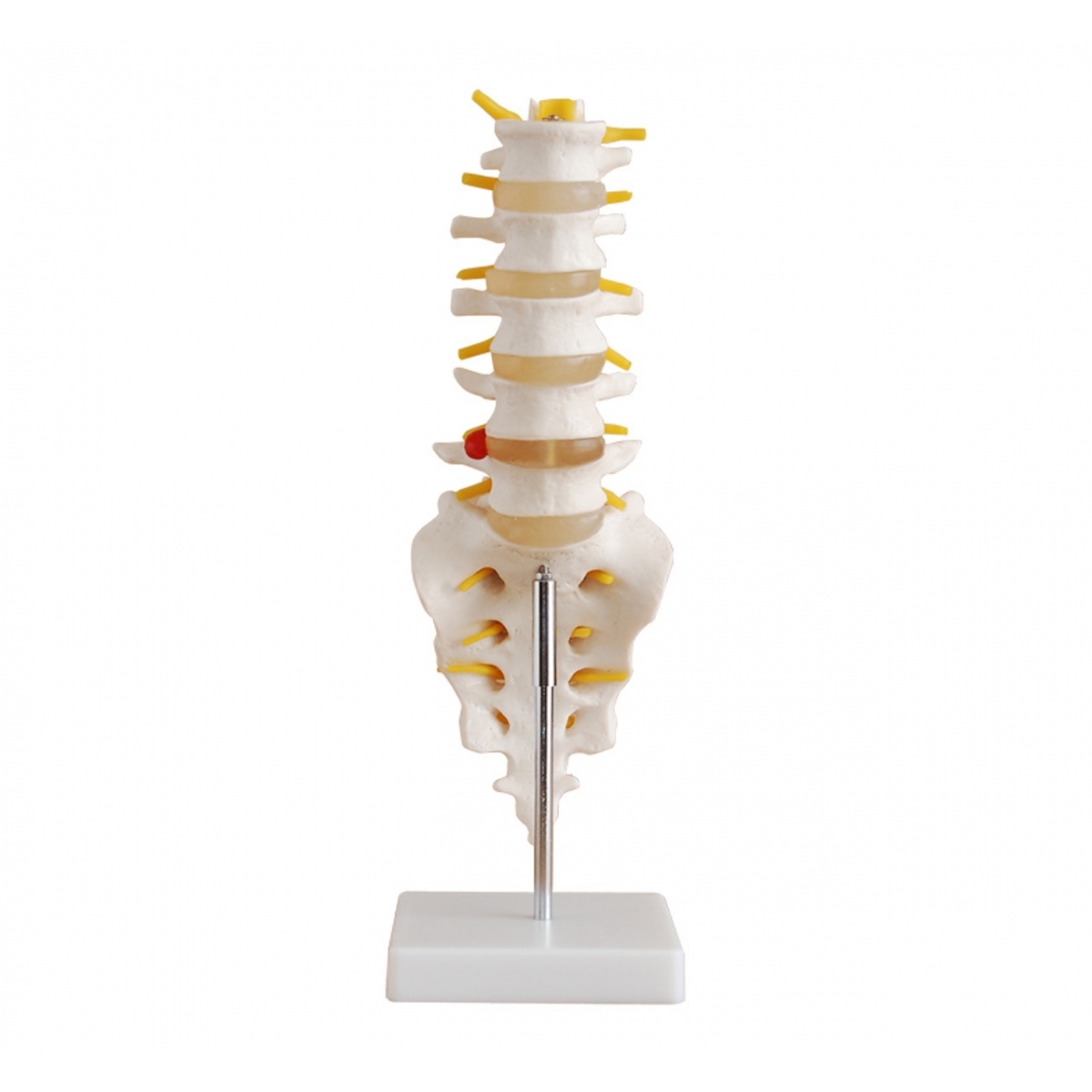
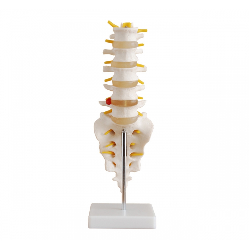
The lumbar spine is where stability, support, and flexibility converge—making it one of the most functionally critical and clinically significant regions in human anatomy. The MYASKRO Life-Size Lumbar Vertebrae Model with Sacrum, Coccyx, and Herniated Disc offers an exceptionally detailed representation of this complex area, tailored for educators, orthopedic specialists, physiotherapists, and anatomy students.
Crafted to show every key component with lifelike accuracy, this model includes five lumbar vertebrae, intervertebral discs, sacrum, coccyx, and realistic yellow spinal nerves. What sets it apart is the clearly visible herniated disc—a powerful visual and tactile tool for demonstrating disc pathology, nerve impingement, and associated back pain symptoms.
Ideal for use in clinical consultations, anatomy labs, chiropractic clinics, or rehabilitation centers, this compact model delivers high educational value in a desk-friendly size. Whether explaining sciatic nerve compression to a patient or helping a student prepare for practical exams, this tool enhances comprehension and improves retention.
Users often ask: “Is it realistic enough to demonstrate pathology effectively?” Without question. The herniation is raised, textured, and positioned at L4-L5 for accurate teaching of one of the most common problem areas in spinal care. Paired with the sacral and coccygeal detail, it gives a full view of lumbosacral biomechanics in a format that’s immediately understandable.
Keywords that match this model's utility include “lumbar spine model,” “herniated disc anatomy,” “life-size spinal nerve demo model,” and “sacrum and coccyx teaching aid.” It serves both search intent and professional intent with equal precision.
“It’s helped me explain disc issues to patients far more effectively. They understand it the moment they see it,” says Dr. Kamal V., Orthopedic Consultant, Delhi.
Simple to display. Easy to use. Clinically valuable. This model isn’t just an object—it’s a daily tool in the hands of those who educate, treat, and care for the spine. Every nerve root and vertebral notch tells a story—and this model helps you tell it well.
The MYASKRO Lumbar Vertebrae Model is engineered to give users a deep, tactile understanding of the lumbar region’s structure and function. From the detailed vertebral bodies to the striking depiction of disc herniation, this model transforms the spine from a theoretical concept into a concrete learning experience.
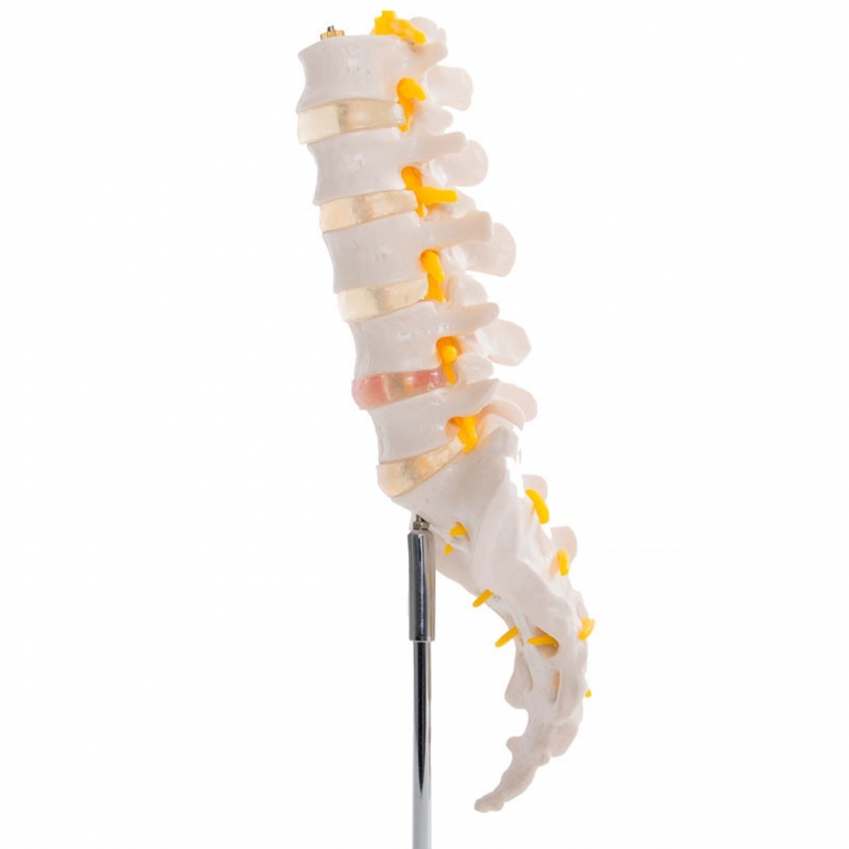
It includes the full lumbar segment (L1–L5), with soft yet durable intervertebral discs separating each vertebra. These discs are clearly defined and slightly compressible, simulating the cushioning role they play in daily movement. The model also features the sacrum and coccyx, adding biomechanical context by showing how the spine transitions into the pelvic region.
One standout detail is the bulging disc at L4-L5. The protrusion is highlighted in red, showing exactly how spinal nerves can become compressed. Instructors can use this to explain sciatic pain, limited mobility, or degenerative disc disease with clarity that’s simply not possible with 2D diagrams.
Wondering about application? It’s ideal for orthopedic instruction, physiotherapy demonstrations, chiropractic education, and neurology consultations. Whether you’re training new professionals or educating a patient about their condition, the model communicates with authority and simplicity.
Built from a high-grade synthetic resin, this model is made to last. It resists chips, warping, and stains—even in high-use environments like classrooms or clinics. The vertebrae are mounted on a stable base with a steel rod, making it easy to rotate and present from any angle.
Semantic keywords like “herniated lumbar disc model,” “nerve root compression teaching aid,” “spinal cord and vertebrae replica,” and “chiropractic lumbar spine tool” all align seamlessly with this model’s purpose and features. It’s optimized not just for anatomy, but for education in action.
“This small model makes a big impact. Patients can visualize their condition, and students gain a physical reference that sticks in memory,” shares Jennifer L., Physiotherapy Lecturer, Birmingham.
When it comes to spinal education, there’s no substitute for something you can point to, rotate, and understand from every angle. That’s exactly what this lumbar model offers—precision, clarity, and the confidence that comes with true anatomical understanding.
When it comes to anatomy models, bigger isn’t always better. What matters is clarity, accuracy, and practical usability. The MYASKRO Lumbar Vertebrae Model with Sacrum, Coccyx, and Herniated Disc proves this point beautifully. It’s compact enough to fit on a desk or consultation table—but detailed enough to explain complex spinal issues with absolute precision.
This model earns its place in any clinical, academic, or therapeutic environment. It supports fast-paced teaching, one-on-one patient education, and even student self-study. Whether you’re guiding a lecture on vertebral alignment or using it to help a patient understand why they’re experiencing lower back pain, the model adapts to your needs with ease.
Its presence elevates credibility. Patients trust what they can see. Students remember what they can touch. And professionals appreciate tools that are as effective as they are durable. With realistic nerve roots, disc textures, and vertebral landmarks, it gives your audience something to engage with—beyond just words.
Frequently asked: “Is it suitable for remote or mobile teaching?” Definitely. Its light weight and stable base mean it travels well, whether across campus or across the clinic. It’s also ideal for pop-up workshops, continuing education programs, and medical demonstrations in non-traditional settings.
This model consistently ranks high for terms like “lumbar spine model with herniated disc,” “sacral vertebrae teaching model,” “life-size lower spine replica,” and “anatomical lumbar model for patient education.” That’s because it hits all the right notes—form, function, and purpose in one smart design.
“It’s one of the most used models in our training center. Explaining back pain, posture, or disc injuries becomes far easier with this on hand,” says Dr. Lee M., Clinical Educator, Singapore.
Great tools simplify complex knowledge—and in spine education, simplicity leads to understanding, and understanding leads to better outcomes. This model does just that. It becomes the kind of product that quietly improves your work, day after day. And when that happens, it’s more than a model—it’s a vital part of how you teach, learn, and heal.
Total Reviews (0)