


 5.8 Feet Tall 96% Anatomical Accuracy Premium Quality | MYASKRO")
₹ 14,699 ₹ 33,039
")
₹ 16,599 ₹ 53,994

₹ 3,999 ₹ 7,315

₹ 1,899 ₹ 2,241
 Anatomical Model")
₹ 3,199 ₹ 6,831
 - MYASKRO")
₹ 25,999 ₹ 40,119

₹ 8,499 ₹ 22,445

₹ 4,199 ₹ 10,555
 With Femur Heads & Pelvis")
₹ 5,499 ₹ 11,617
")
₹ 7,799 ₹ 12,845
|MYASKRO")
₹ 9,299 ₹ 14,545

₹ 11,799 ₹ 18,945
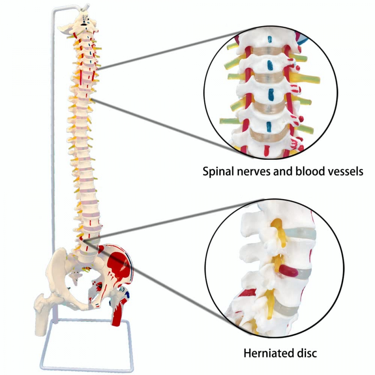
When it comes to understanding the human spine, movement matters. The MYASKRO Flexible Spine Model offers a dynamic and hands-on view of vertebral function, complete with painted muscle origins and insertions, movable femur heads, visible spinal nerves, and a detailed pelvis with occipital plate. It’s more than a teaching aid—it’s a functional simulation of spinal motion and muscular attachment.
Designed for medical students, physiotherapists, orthopedic specialists, chiropractors, and fitness educators, this model allows learners to examine how vertebrae align, how muscles act on bone, and how flexibility, posture, and nerve pathways all work together in the spine’s operation. The entire spinal column—from cervical to coccygeal—is represented with life-like curvature and movement, and the model flexes naturally to replicate bending, twisting, and real-world spinal motion.
Key features include:
For student labs, the model reinforces textbook learning by giving learners a tangible, mobile frame to manipulate and explore. For clinical professionals, it becomes an indispensable patient education tool—illustrating the mechanics of back pain, disc herniation, postural misalignment, and muscular strain with real-time movement and reference points.
Mounted securely on a supportive stand, the model remains upright for display but detaches easily for hands-on demonstrations. Its durable materials and anatomical fidelity make it suitable for daily handling, in-depth instruction, and detailed biomechanical discussion.
As one chiropractor shared, “This model lets me show patients what their spine is doing—and why it matters. It turns anatomy into action.”
Whether in a lecture hall or clinical setting, the MYASKRO Flexible Spine Model brings anatomy to life—one vertebra, one nerve, one muscle at a time.
The MYASKRO Flexible Spine Model delivers the trifecta of functional anatomy—skeletal structure, muscular attachment, and neural integration—all with interactive realism. This model is engineered not only to demonstrate static anatomy, but to give learners and practitioners a clear, hands-on understanding of how the spine moves, how muscles pull, and how nerves flow through it all.
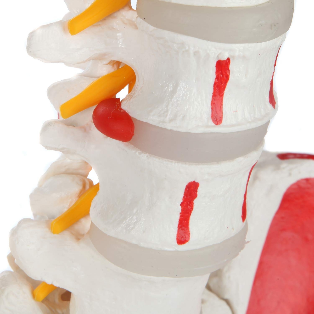
What sets this model apart is its full flexibility. You can bend, rotate, and manipulate the spine to show cervical extension, thoracic kyphosis, lumbar lordosis, or sacral curvature. This flexibility is particularly useful when teaching spinal mechanics, postural correction, or injury patterns such as disc herniation or nerve impingement.
Every detail is carefully designed for educational depth:
This model becomes especially powerful in environments where active demonstration is key:
The flexibility also allows educators to reinforce concepts like spinal segment mobility, intervertebral disc function, and the effects of movement restriction. By observing how the model flexes or resists motion, learners build intuitive understanding—not just memorized terms.
Constructed from high-quality, medical-grade materials, the model is built for years of repeated use. The flexible column holds shape while allowing smooth articulation, and the colored markers stay vivid after countless demonstrations.
As one physiotherapy lecturer said, “It’s not just a model—it’s a conversation starter. Students suddenly ‘see’ what the spine is doing beneath the skin.”
With this model, you don’t just study the spine. You demonstrate it. You move it. You teach it in motion.
The spine isn’t static. It bends, rotates, compresses, and responds to every movement the body makes. That’s why a rigid model can only teach part of the story. The MYASKRO Flexible Spine Model completes that story—offering full anatomical clarity with the added benefit of real-life flexibility, visible nerve pathways, and color-coded muscle functions.
This isn’t just a model for identifying vertebrae. It’s a tool for exploring how movement happens, how alignment affects function, and how disruptions in posture or structure can lead to discomfort or dysfunction. Every demonstration becomes a dynamic lesson—whether it’s showing students how the lumbar spine reacts during flexion, or explaining to a patient why nerve compression in the cervical spine causes symptoms in the arm.
Its painted red (origin) and blue (insertion) muscle markers instantly communicate muscular leverage and directional force. Learners don’t just memorize attachment points—they visualize how muscles create motion. The visible nerve roots enhance this further by tying structure to neurology, showing how misalignments or disc issues can lead to real clinical symptoms. It transforms rote memorization into clinical insight.
Educators in all fields are finding the model indispensable:
The included pelvis and removable femur heads create a complete lumbopelvic view, allowing educators to discuss core stability, sacroiliac dysfunction, and hip-spine interactions. And the flexible yet stable construction makes it perfect for repeated use without warping or loosening over time.
As one medical professor noted, “Our students finally grasped spinal mechanics the moment they moved this model. It connects the dots in ways a lecture never could.”
With the MYASKRO Flexible Spine Model, learning is no longer limited to static diagrams. You can now see the motion, feel the interaction, and teach the spine the way the human body actually experiences it—in motion, in balance, and in context.
Total Reviews (0)