 | Myaskro")
₹ 17,499 ₹ 28,945

₹ 18,999 ₹ 31,845
Overview
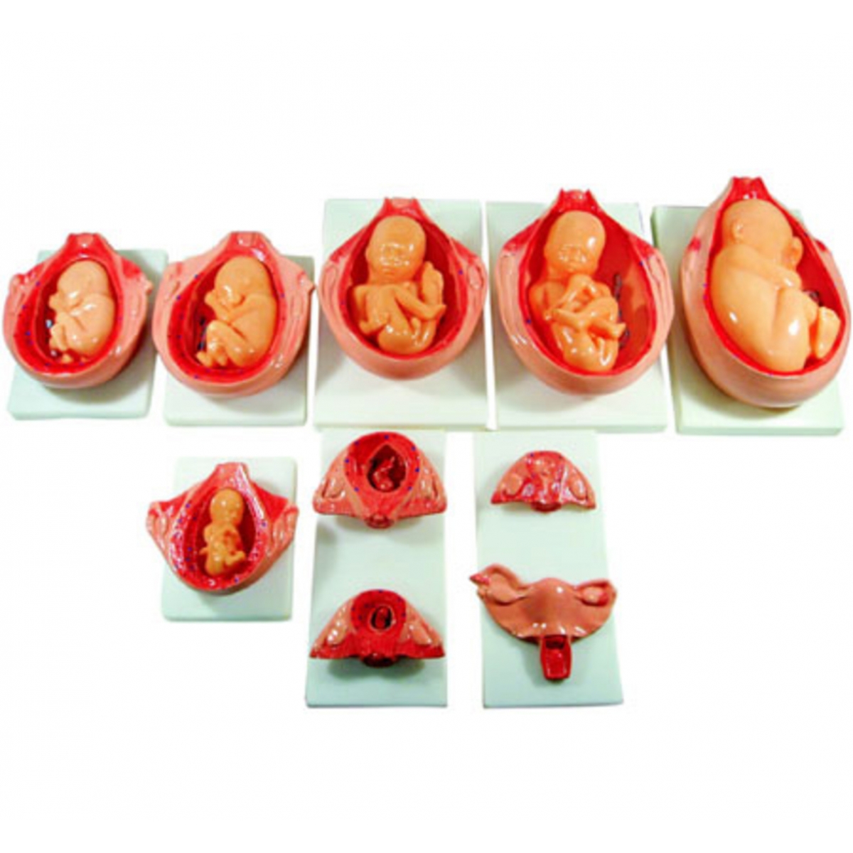
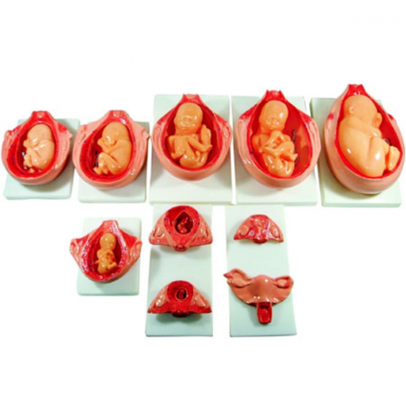
Ten mounted models tracing human development from early embryo/implantation to near-term fetus. Each board shows a sectioned gravid uterus with amniotic cavity, chorion/amnion, placenta and cord, and fetus in situ, allowing clear visualization of uterine growth, placental maturation, and changing fetal lie/presentation. Instructional aid; not a medical device.
Implantation in decidua (chorionic villi beginning).
Early embryo with yolk sac; amniotic cavity formation.
Early fetus with limb buds; primitive placenta.
4–8. Progressive fetal growth across the second trimester—ossifying skeleton, facial development, external genitalia, increasing vernix/lanugo, and enlarging placenta (chorion frondosum/decidua basalis).
9–10. Late-trimester fetus with flexed posture, cephalic presentation; mature placenta and coiled umbilical cord.
Teaching objectives
Differentiate embryonic vs fetal period and correlate with organogenesis and growth.
Identify the placenta, membranes (amnion/chorion), yolk sac, and umbilical vessels; discuss maternal–fetal exchange.
Demonstrate uterine enlargement, cervical changes, and fetal positions (breech vs cephalic for counselling).
Explain common obstetric pathology in context: placenta previa/accreta (concepts), oligohydramnios/polyhydramnios, and IUGR vs normal growth curves.
Support antenatal classes, midwifery teaching, OB-GYN clinics, and OSCE stations.
Set of 10 Medical Grade PVC relief models on wipe-clean boards; high-contrast, hand-painted detail.
Wall-mount or tabletop display; numbered legends included.
Care: wipe with mild detergent or 70% alcohol; avoid solvents/heat.
Ideal users: Obstetrics & gynecology departments, nursing/midwifery colleges, antenatal educators, public-health displays.
Total Reviews (0)