

₹ 1,799 ₹ 4,301

₹ 4,799 ₹ 9,316

₹ 38,999 ₹ 45,645
The miracle of prenatal development is often hidden from view—complex, layered, and essential. The MYASKRO Fetus with Viscus & Placenta Anatomical Model changes that by providing a vivid, three-dimensional exploration of fetal anatomy, organ placement, and maternal-fetal connection via the placenta. It’s a complete prenatal teaching tool in one compact, precise display.
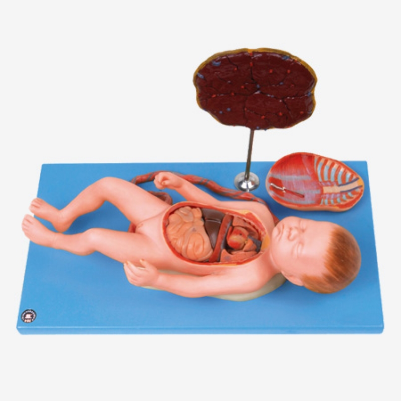
This detailed model features a life-sized fetus with removable internal organs, an umbilical cord, and a segmented placenta with attached membranes. Whether you’re training medical students in obstetrics, teaching midwifery students about fetal development, or guiding expecting parents through childbirth education—this model delivers anatomy that’s both accessible and accurate.
The visceral cavity reveals essential organs like the intestines, liver, heart, and lungs, all in realistic proportion and orientation. The chest plate can be removed for direct visualization of internal fetal structures, while the umbilical cord and placenta demonstrate the critical link between mother and child. It’s a hands-on, tactile way to show where life begins—and how it’s sustained before birth.
Key Features:
This model is ideal for:
As one anatomy instructor put it, “It’s the most complete fetal model I’ve used—it gives students a full visual language for understanding life before delivery.”
For any setting where fetal anatomy is taught, explained, or appreciated, the MYASKRO model brings function, form, and clarity into one elegant display.
The MYASKRO Fetus with Viscus & Placenta Anatomical Model is more than a teaching aid—it's a three-dimensional journey into prenatal life. From the precise arrangement of internal organs to the critical pathways of maternal-fetal exchange, every component of this model is designed to make complex prenatal anatomy intuitive and visible.
Unlike flat illustrations or animated videos, this physical model allows learners to explore fetal development tactilely and spatially. Students can open the thoracoabdominal cavity, examine the fetal liver, intestines, heart, and lungs, and understand how each organ forms and functions at this early stage. The removable chest plate allows repeated, risk-free exploration—ideal for both structured learning and self-guided study.
Attached to the fetal model is an umbilical cord that leads to a lifelike placenta and membranes, providing an anatomical basis for understanding fetal circulation and the exchange of nutrients, oxygen, and waste. The vascular tree on the placenta is realistically colored and textured, allowing clear differentiation between maternal and fetal contributions. This is particularly important when teaching about:
Medical and midwifery students benefit from this model by being able to visualize what would otherwise remain abstract. It also plays a critical role in antenatal education, helping expecting parents understand the connection between mother and baby and reinforcing trust in care providers by making the invisible, visible.
Built on a sturdy, cleanable base, this model is perfectly suited for:
Its durable PVC construction ensures it withstands frequent handling, while the lifelike scale ensures realism without overwhelming classroom space. And because each structure is individually molded and painted, detail is never sacrificed for compactness.
As one midwife trainer put it, “You can teach an entire trimester's worth of anatomy with this one model. It’s complete, clear, and surprisingly emotional to show in class.”
For anyone seeking to deepen understanding of fetal life, this model becomes not just a tool—but a bridge between textbook knowledge and real-world insight.
The MYASKRO Fetus with Viscus & Placenta Anatomical Model stands apart as a tool of both precision and empathy. It doesn’t just show what a fetus looks like—it reveals how life unfolds before birth. Every structure, from the visible viscera to the vascularized placenta, is crafted to offer not only anatomical clarity but also contextual understanding of how the human body develops, thrives, and prepares for entry into the world.
For medical and nursing students, this model is a valuable asset in courses that cover embryology, pediatrics, obstetrics, or neonatology. With its removable internal components, it makes it easy to explore complex systems—respiratory, digestive, circulatory—before they fully mature. It becomes a platform for asking deeper questions: How does fetal blood bypass the lungs? Why does the liver appear so prominent at this stage? What happens if organ development is interrupted?
Midwifery instructors use this model to demonstrate fetal positioning and organ development across trimesters. In antenatal classes, it allows parents to grasp how their baby grows—not just in size, but in function and form. For doulas, childbirth educators, and OB-GYNs, it becomes a tool of trust—visually explaining what’s happening inside a womb and demystifying the journey of pregnancy.
Because it’s mounted on a stable base and built from durable, easy-to-clean PVC, the model holds up to repeated use in both academic and clinical environments. Its compact footprint allows for tabletop demonstration or permanent display, while the lifelike color and touch make it engaging and approachable for learners of all backgrounds.
Its educational impact is well-documented. As one fetal medicine specialist shared, “This model has changed how we teach. Students retain more when they interact physically with a full, cross-sectional fetus rather than just read about it. It brings clarity, and with it, confidence.”
And that’s what truly sets this model apart—it brings confidence. Confidence to teach. Confidence to learn. Confidence to explain life’s most formative stage with accuracy, compassion, and clarity.
For anyone involved in prenatal care, medical education, or anatomical instruction, this isn’t just a model—it’s a foundational investment in understanding the human story from its very beginning.
Total Reviews (0)