


₹ 6,899 ₹ 17,530

₹ 5,199 ₹ 7,290
 With Special Lamination")
₹ 1,599 ₹ 3,865
 With Special Lamination")
₹ 1,599 ₹ 3,865

₹ 15,499 ₹ 22,379

₹ 8,999 ₹ 12,865
Components
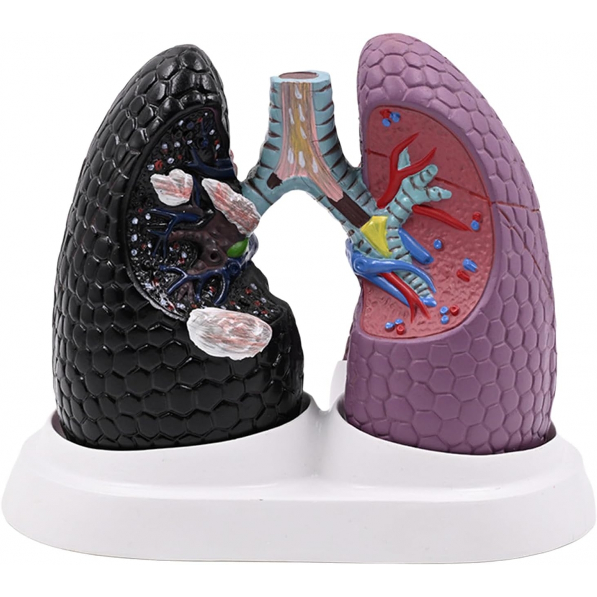
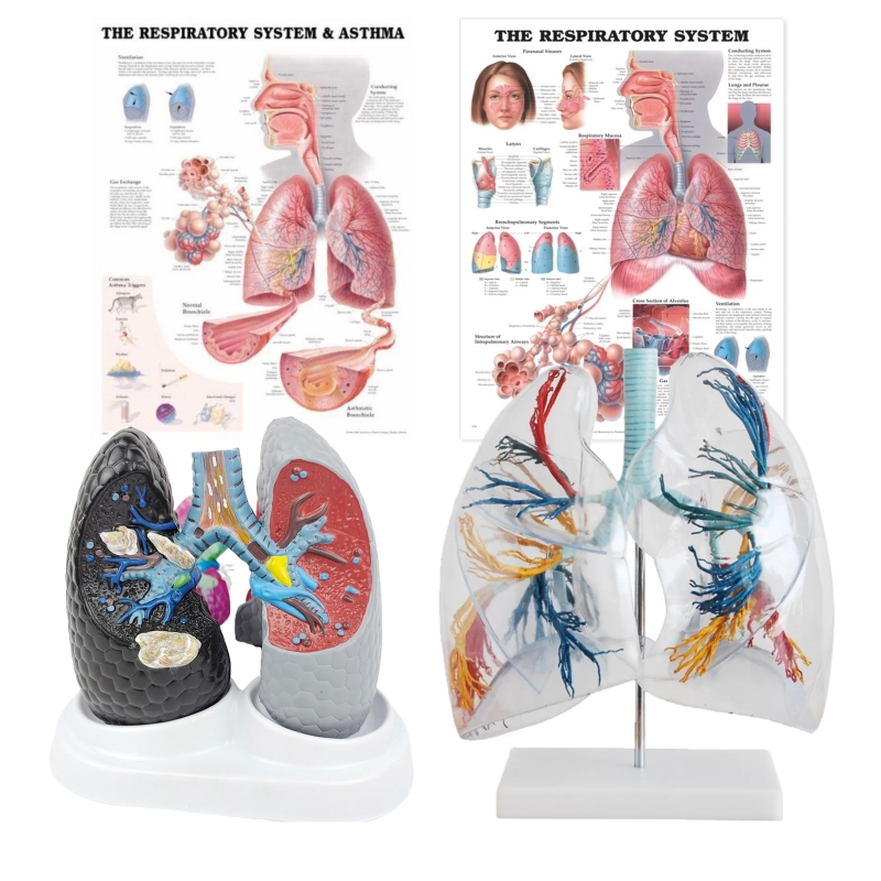
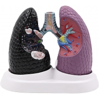
Health & Pathological Lungs Model: Bilateral lungs with trachea–bronchial tree and cut-sections demonstrating representative pathology (airway hyperreactivity/asthma with bronchospasm and mucus plugging, chronic bronchitis, emphysema with alveolar septal loss, focal consolidation, neoplastic lesion/“smoker’s lung”).
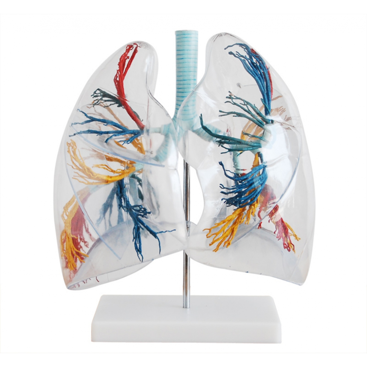
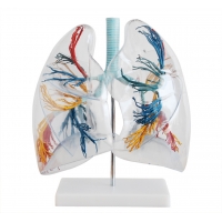
Transparent Lungs Segment Model: Clear lobar shells with color-coded bronchopulmonary segments (segmental bronchi and paired pulmonary arterial branches) for 3-D segmental orientation; visible oblique and horizontal fissures.
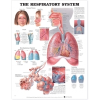
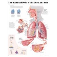
Charts (52 × 70 cm, laminated with rollers):
Respiratory System (airways, ventilation, gas exchange)
Respiratory System & Asthma (pathophysiology, triggers, airway remodeling)
Learning objectives
Identify lobes, fissures, and 20 bronchopulmonary segments; correlate segments with segmentectomy and targeted physiotherapy.
Explain airflow limitation in asthma/COPD (bronchospasm, mucosal edema, mucus hypersecretion, loss of elastic recoil).
Demonstrate alveolar–capillary interface and sites of consolidation/atelectasis on sectional surfaces.
Map symptoms to anatomy: wheeze (bronchial narrowing), hypoxemia (V/Q mismatch), hemoptysis (tumor/bronchitis).
Use charts to teach ventilation mechanics, mucociliary clearance, and asthma education for patients/guardians.
Specifications
Models: Non-Toxic Medical Grade PVC.
Charts: heavy-gauge lamination; dry-wipe compatible; roller bars for mounting.
Cleaning: mild detergent or 70% alcohol wipe; avoid solvents/heat.
Intended use: instructional aids for UG/PG medicine, pulmonology, thoracic surgery, anesthesia, physiotherapy, nursing; OSCE/skill-lab stations.
Total Reviews (0)