

₹ 1,999 ₹ 5,245

₹ 4,799 ₹ 18,407

₹ 51,999 ₹ 105,557

₹ 103,899 ₹ 199,343

₹ 2,899 ₹ 4,537

₹ 3,599 ₹ 6,712
 - Myaskro")
₹ 148,699 ₹ 293,767

₹ 38,999 ₹ 45,645
 | Myaskro")
₹ 17,499 ₹ 28,945

₹ 29,499 ₹ 58,876

₹ 18,999 ₹ 31,845
Overview
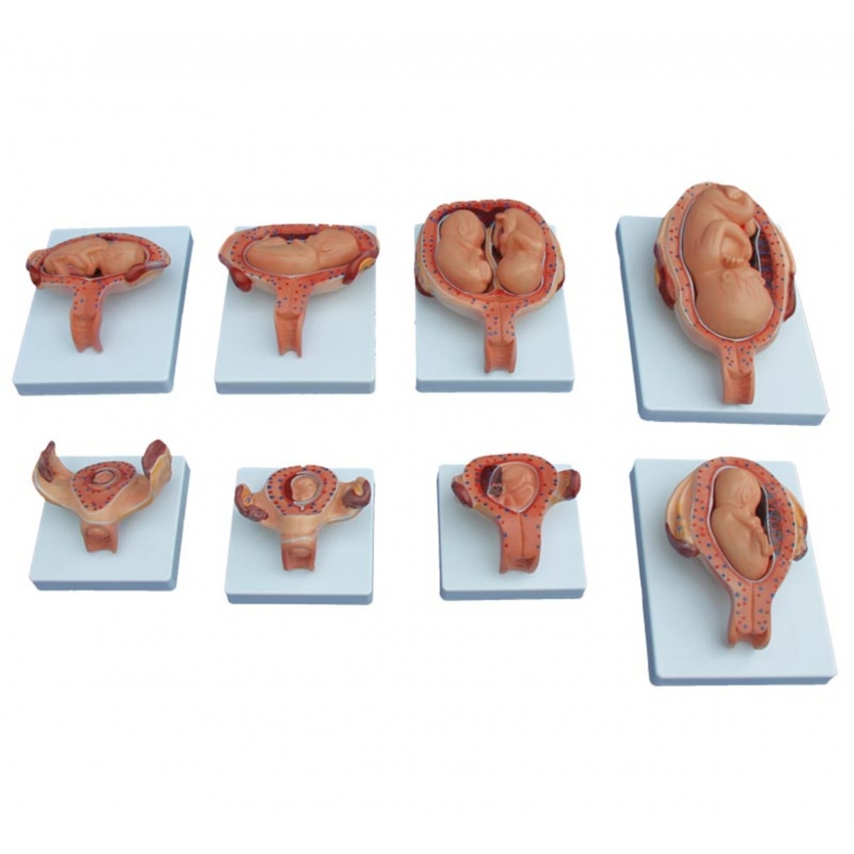
Eight mounted uterine cross-sections depicting normal human development from implantation to late gestation. Each plate shows the uterus, placenta, membranes (amnion/chorion), umbilical cord, and fetus in situ; one plate illustrates twin gestation. Instructional aid; not a medical device.
Implantation/chorionic villi forming in decidua.
Early embryo with yolk sac and amniotic cavity.
Early fetus with limb buds; primitive placenta.
First-trimester fetus with enlarging amnion and cord.
Second-trimester growth—proportional limb and craniofacial development.
Twin pregnancy (schematic, for comparison).
Third-trimester fetus—increasing uterine size and placental maturation.
Late pregnancy/cephalic lie—reduced free space, mature placenta and membranes.
Distinguish embryonic vs fetal periods; correlate with organogenesis and somatic growth.
Identify the placenta, chorion, amnion, yolk sac, and umbilical vessels; explain maternal–fetal exchange.
Demonstrate uterine enlargement, cervical relations, and fetal lie/presentation for antenatal counselling.
Discuss normal vs multiple gestation and introduce common clinical concepts (IUGR vs normal growth, fluid volume changes).
8 relief plates, Medical Grade PVC, hand-painted, wipe-clean.
Stable backboards for tabletop or wall display; numbered labels with legend card.
Cleaning: mild detergent or 70% isopropyl alcohol; avoid solvents/heat.
Ideal users: Obstetrics & gynaecology departments, nursing/midwifery colleges, antenatal educators, public-health exhibits, OSCE stations.
Total Reviews (0)