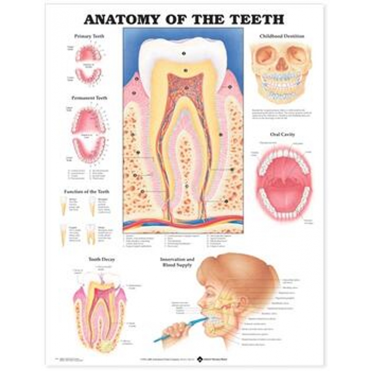
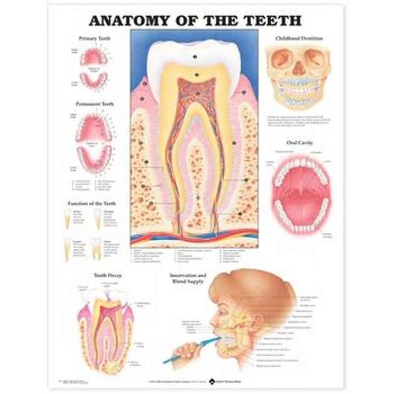
Anatomy of the Teeth Chart (52x70 cm) with Special Lamination
This high-quality anatomical chart clearly illustrates the structure and function of human teeth, including primary and permanent dentition, nerve supply, blood vessels, dental cavities, and more. Perfect for MBBS students, dental colleges, clinics, and medical classrooms, this chart is both informative and display-friendly.
Key Features
- Detailed dental anatomy: Includes sections on tooth decay, oral cavity, childhood dentition, innervation, and blood supply
- Lamination: Prevents tearing, allows easy cleaning, and increases lifespan
- Large size: 52 x 70 cm for enhanced readability and presentation
- Color-coded illustrations: Designed for quick understanding and visual clarity
Perfect For
- MBBS and dental students studying oral anatomy
- Medical and dental colleges
- Dental clinics for patient education
- Hospitals, classrooms, and health centers
Specifications
- Size: 52 x 70 cm
- Material: Heavy-duty laminated poster
- Finish: Glossy lamination for long-term use and easy cleaning
Frequently Asked Questions
Is this chart suitable for dental students?
Yes, it’s designed for dental and MBBS students to understand tooth anatomy, including nerve and blood supply.
Does it come laminated?
Yes, it has a special lamination that makes it durable and easy to clean with a dry cloth.
This chart is more than a poster — it’s a complete teaching aid for oral anatomy, made to last and impress in any medical learning environment.
")