

₹ 8,299 ₹ 12,965
")
₹ 8,799 ₹ 18,945
")
₹ 3,999 ₹ 8,965
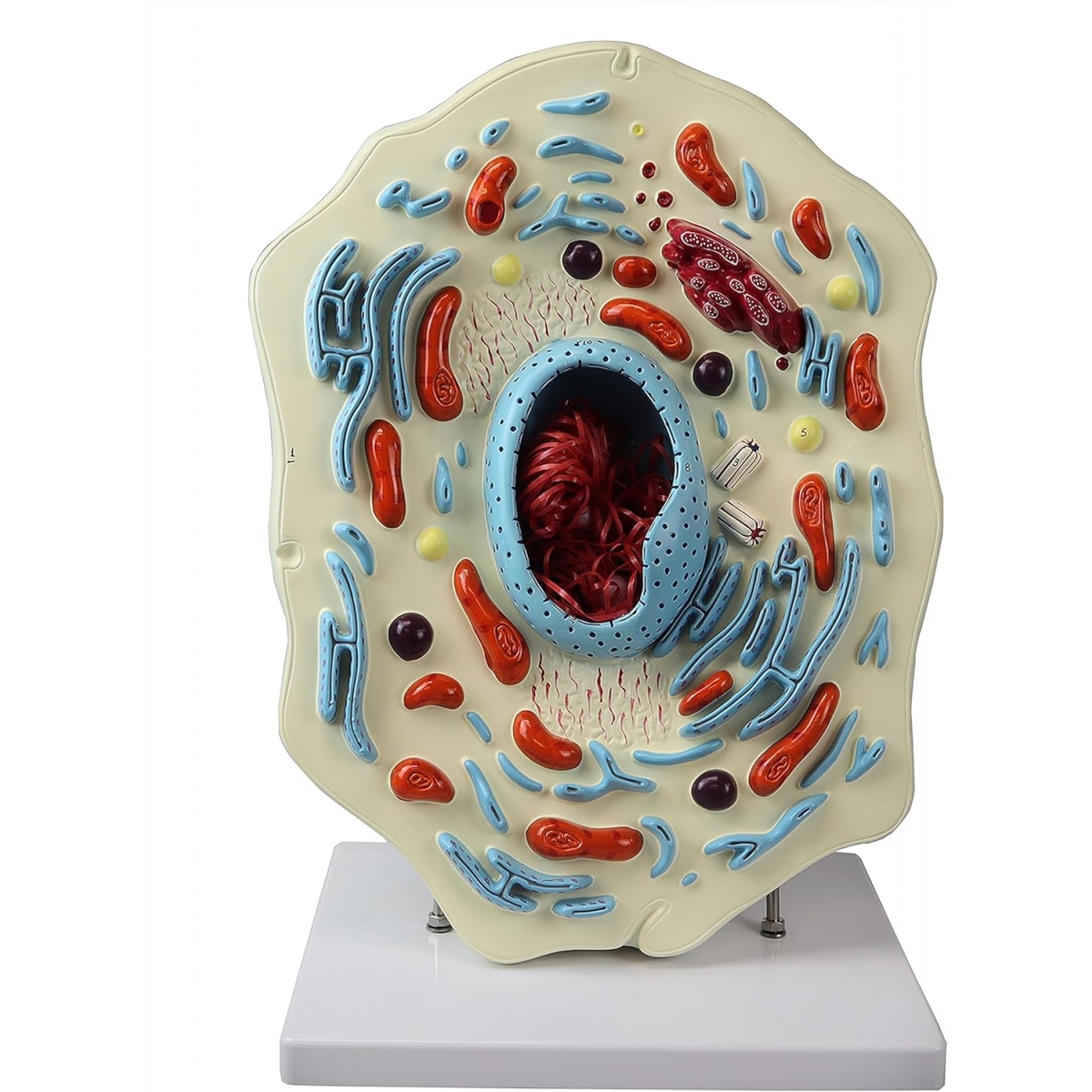
Giant 3D Animal Cell Model (on Stand)
High-visibility teaching model showing a sectioned eukaryotic animal cell with raised, color-coded organelles for quick identification and explanation. Ideal for viva prep, classroom demos and patient/parent education in clinics.
What you’ll study (with brief lay cues):
Nucleus with chromatin & nucleolus – the cell’s control center (DNA & ribosome assembly).
Nuclear envelope with pores – gated transport to/from cytoplasm.
Rough & Smooth Endoplasmic Reticulum – protein synthesis (ribosome-studded) and lipid/drug metabolism.
Golgi apparatus with vesicles – protein modification, packaging and trafficking.
Mitochondria – ATP production (cell “powerhouses”).
Lysosomes/Peroxisomes – digestion and detox.
Centrosome with centrioles – spindle organization for cell division.
Cytoplasm & plasma membrane – fluid matrix and selective barrier.
Built for teaching
Giant, relief-sculpted layout for clear viewing from the back row.
Crisp color coding and numbered landmarks for quizzes.
Durable, wipe-clean PVC; mounted on a stable base.
Perfect for MBBS/BDS, nursing, biology labs, schools and skills centers.
Total Reviews (0)